. 2019 Nov;33(11):12164-12174.
doi: 10.1096/fj.201900992R. Epub 2019 Aug 1.

TREM2 deficiency aggravates α-synuclein-induced neurodegeneration and neuroinflammation in Parkinson's disease models

Affiliations

TREM2 deficiency aggravates α-synuclein-induced neurodegeneration and neuroinflammation in Parkinson's disease models

Ying Guo et al. FASEB J. 2019 Nov.

Abstract

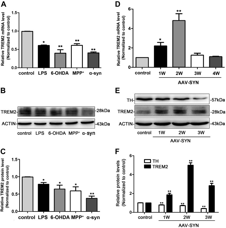
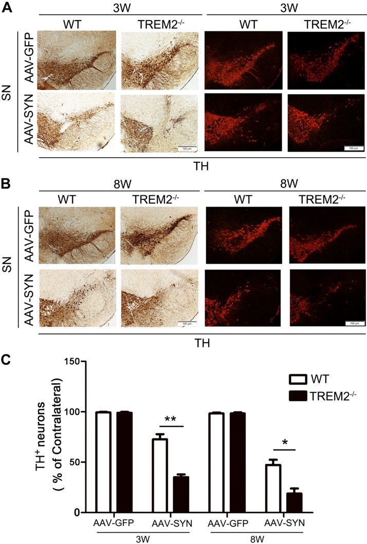
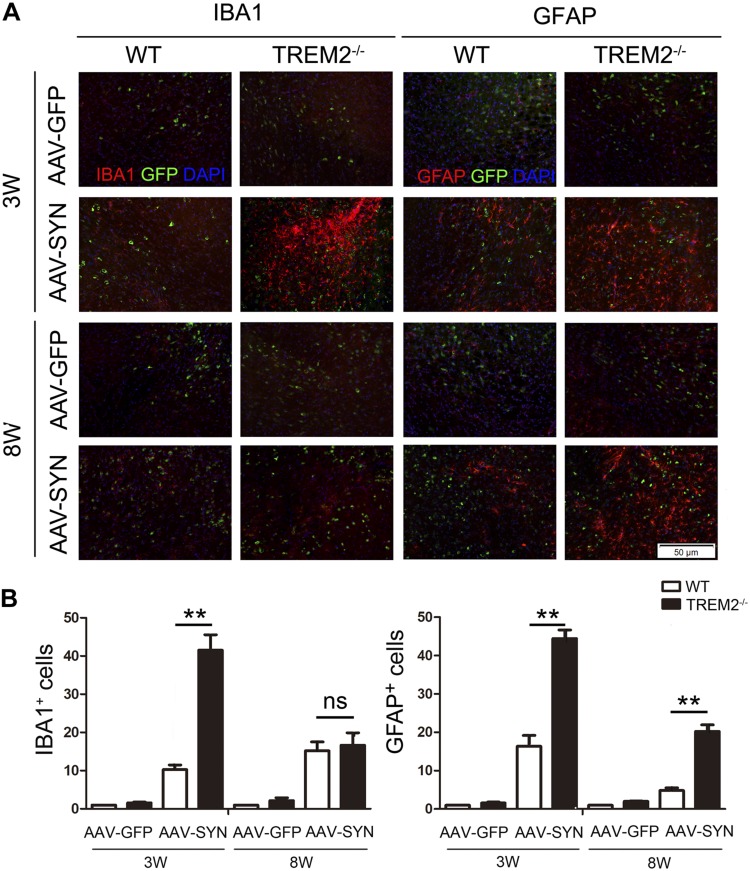
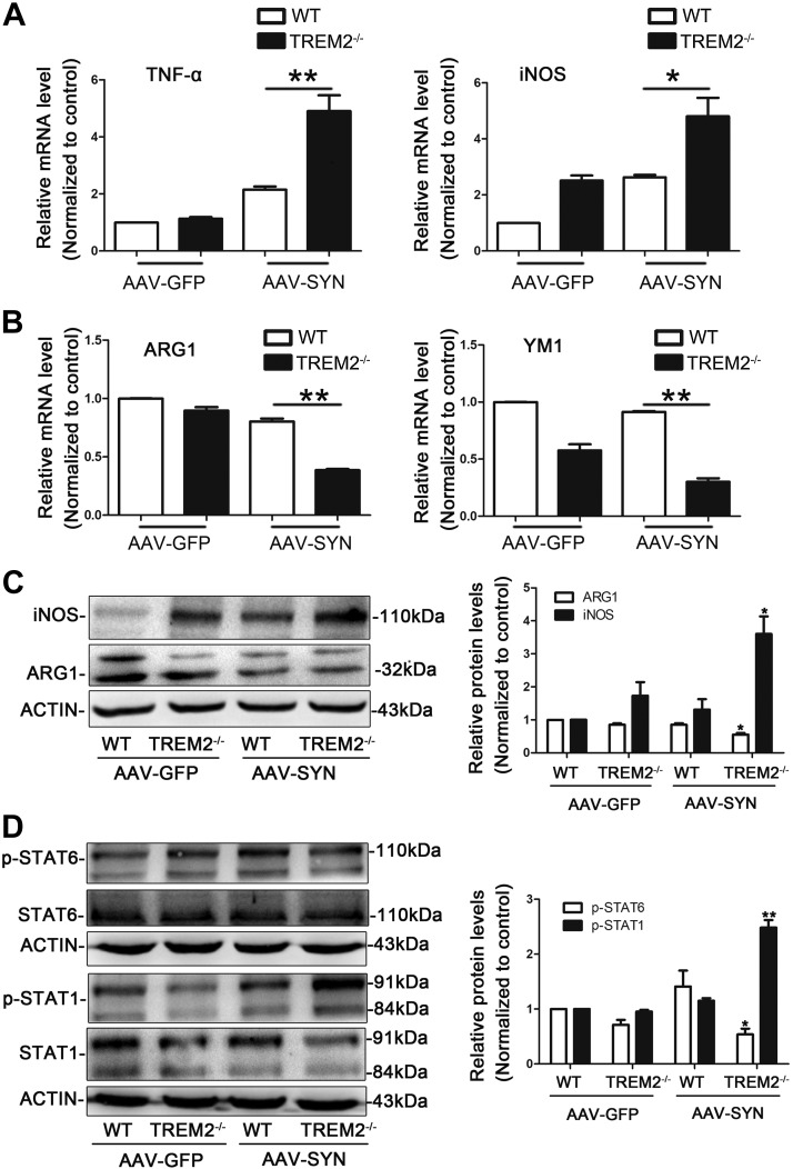
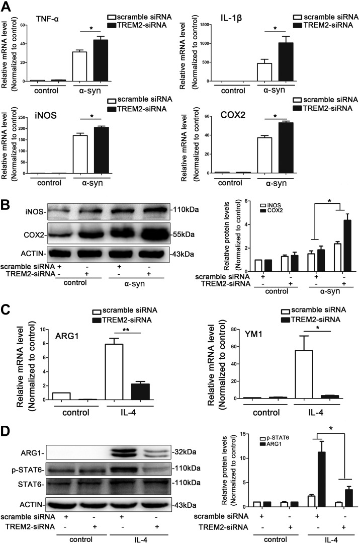
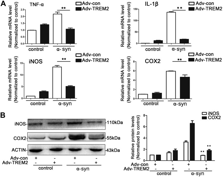
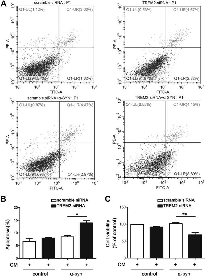
Variants in the gene encoding the triggering receptor expressed on myeloid cells 2 (TREM2) are known to increase the risk of developing Alzheimer disease and Parkinson's disease (PD). However, the potential role of TREM2 effect on synucleinopathy has not been characterized. In this study, we investigated whether loss of TREM2 function affects α-synucleinopathy both in vitro and in vivo. In vitro, BV2 microglial cells were exposed to α-synuclein (α-syn) in the presence or absence of TREM2 small interference RNA. For in vivo studies, wild-type controls and TREM2 gene knockout mice were intracranially injected in the substantia nigra with adeno-associated viral vectors expressing human α-syn (AAV-SYN) to induce PD. Our results revealed that knockdown of TREM2 aggravated α-syn-induced inflammatory responses in BV2 cells and caused greater apoptosis in SH-SY5Y cells treated with BV2-conditioned medium. In mice, TREM2 knockout exacerbated dopaminergic neuron loss in response to AAV-SYN. Moreover, both in vitro and in vivo TREM2 deficiency induced a shift from an anti-inflammatory toward a proinflammatory activation status of microglia. These data suggest that impairing microglial TREM2 signaling aggravates proinflammatory responses to α-syn and exacerbates α-syn-induced neurodegeneration by modulating microglial activation state.-Guo, Y., Wei, X., Yan, H., Qin, Y., Yan, S., Liu, J., Zhao, Y., Jiang, F., Lou, H. TREM2 deficiency aggravates α-synuclein-induced neurodegeneration and neuroinflammation in Parkinson's disease models.

Keywords: PD; inflammation; microglia; α-syn.


Conflict of interest statement

The authors are thankful to Dr. Ruth G. Perez (Texas Tech University Health Science Center, El Paso, TX, USA) for editorial assistance with the manuscript. This work was supported by grants from the Shandong Natural Science Foundation (ZR2018MH043) and the Shandong Province Science and Technology Program (2017GSF18187). The authors declare no conflicts of interest.

Figures

Figure 1
Figure 1
The expression pattern of TREM2 in BV2 microglial cells and AAV-SYN–induced PD mice models. A) BV2 microglial cells were exposed to LPS (100 ng/ml), MPP+ (50 μM), 6-OHDA (75 μM), and α-syn (500 nM) for 24 h; the mRNA level of TREM2 was analyzed with real-time RT-PCR. B, C) Representative immunoblot documents and summarized data showing the protein level of TREM2 in BV2 microglial cells after being exposed to LPS (100 ng/ml), MPP+ (50 μM), 6-OHDA (75 μM), and α-syn (500 nM) for 24 h. D) Real-time RT-PCR analysis of TREM2 mRNA level in the SN at different time points after AAV-SYN injection in mice. E, F) Representative immunoblot documents and summarized data showing the protein levels of TREM2 and TH in the striatum at different time points after AVV-SYN injection in mice. All data are presented as the mean ± sem of triplicate independent experiments. *P < 0.05, **P < 0.01 vs. control.
Figure 2
Figure 2
TREM2 knockout aggravates AAV-SYN–associated neurodegeneration in vivo. AAV-GFP and AAV-SYN were unilaterally injected in the SN of WT and TREM2−/− mice. Brains were collected at 3 and 8 wk after virus injection; immunohistochemical and immunofluorescent staining for TH were performed in the SN. A, B) Representative image of TH immunoreactive neurons in the injected SN at 3 and 8 wk after virus injection. C) Quantification of TH+ neurons in the SN of WT and TREM2−/− mice using unbiased stereology at 3 and 8 wk after virus injection. Data are presented as means ± sem, n = 4–6 mice/group. *P < 0.05, **P < 0.01.
Figure 3
Figure 3
TREM2 knockout exacerbates AAV-SYN–induced microgliosis and astrogliosis in vivo. AAV-GFP and AAV-SYN were unilaterally injected in the SN of WT and TREM2−/− mice. Mice were euthanized at 3 and 8 wk after virus injection. A) Immunofluorescence for IBA1-positive microglia and GFAP-positive astrocytes (red) in ipsilateral side of the SN. B) Quantification of immunofluorescence data showed that TREM2 knockout exacerbated AAV-SYN–induced microgliosis and astrogliosis. ns, no significance. Data are presented as means ± sem, n = 6 mice/group. **P < 0.01.
Figure 4
Figure 4
TREM2 knockout induces a shift from anti- to proinflammatory microglial activation state in AAV-SYN PD mice. AAV-GFP and AAV-SYN were unilaterally injected in the SN of WT and TREM2−/− mice. Mice were euthanized at 3 wk after virus injection. A, B) The expression of the proinflammatory marker TNF-α, iNOS (A), and anti-inflammatory marker ARG1, YM1 (B) in the ipsilateral SN were detected by real-time RT-PCR. *P < 0.05, **P < 0.01. C) Representative immunoblot and quantitative analysis of iNOS and ARG1 protein levels in the ipsilateral SN of WT and TREM2−/− mice. D) Representative immunoblot and quantitative analysis of p-STAT1 and p-STAT6 protein levels in the ipsilateral SN of WT and TREM2−/− mice. Data are presented as means ± sem, n = 4 animals/group. *P < 0.05, **P < 0.01 vs. WT + AAV-SYN group.
Figure 5
Figure 5
TREM2 deficiency leads to enhanced proinflammatory gene expression in BV2 microglial cells. A, B) TREM2 knockdown increased the expression of proinflammatory markers. Relative mRNA levels of TNF-α, IL-1β, iNOS, and COX2 were detected by real-time RT-PCR in control and TREM2 siRNA-treated BV2 cells after exposure to α-syn for 4 h (A). Representative immunoblot documents and summarized data showing the protein levels of iNOS and COX2 in control and TREM2 siRNA-treated BV2 cells after exposure to α-syn for 4 h (B). C, D) TREM2 knockdown decreased the expression of related anti-inflammatory markers in BV2 cells. Relative mRNA levels of ARG1 and YM1 were detected by real-time RT-PCR in control and TREM2 siRNA-treated BV2 cells after exposure to IL-4 for 24 h (C). The protein levels of ARG1 and p-STAT6 in control and TREM2 siRNA-treated BV2 cells after exposure to IL-4 for 24 h were detected by Western blot analysis (D). Data are presented as the mean ± sem of triplicate independent experiments. *P < 0.05, **P < 0.01.
Figure 6
Figure 6
TREM2 overexpression suppresses α-syn–induced proinflammatory responses in BV2 microglial cells. Cells were transfected with TREM2 vector or a control vector for 48 h, followed by treatment with α-syn for another 4 h. Cells were then collected for the following analysis. A) mRNA levels of TNF-α, IL-1β, COX-2, and iNOS were measured by real-time RT-PCR. **P < 0.01. B) Protein levels of COX-2 and iNOS were detected by Western blot analysis. All data are presented as the mean ± sem of triplicate independent experiments. **P < 0.01 vs. adenoviral particles encoding control vectors + α-syn group.
Figure 7
Figure 7
Suppression of TREM2 in microglia is detrimental to cultured neurons. The control and TREM2-knockdown BV2 cells were treated with α-syn for 4 h. CM was then harvested and added into SH-SY5Y neuron culture for another 24 h. A, B) The apoptosis of SH-SY5Y cells were assessed by Annexin V-PI staining using flow cytometry (A), and the apoptotic rates were shown in graph (B). C) The cell viability of SH-SY5Y cells were detected using MTT assay. Data are presented as the mean ± sem of triplicate independent experiments. *P < 0.05, **P < 0.01.

References

    1. Dickson D. W. (2018) Neuropathology of Parkinson disease. Parkinsonism Relat. Disord. 46 (Suppl 1), S30–S33 - PMC - PubMed
    1. Perry V. H. (2012) Innate inflammation in Parkinson’s disease. Cold Spring Harb. Perspect. Med. 2, a009373 - PMC - PubMed
    1. Jha M. K., Lee W. H., Suk K. (2016) Functional polarization of neuroglia: implications in neuroinflammation and neurological disorders. Biochem. Pharmacol. 103, 1–16 - PubMed
    1. Moehle M. S., West A. B. (2015) M1 and M2 immune activation in Parkinson’s disease: foe and ally? Neuroscience 302, 59–73 - PMC - PubMed
    1. Le W., Wu J., Tang Y. (2016) Protective microglia and their regulation in Parkinson’s disease. Front. Mol. Neurosci. 9, 89 - PMC - PubMed

Publication types

LinkOut - more resources