. 2019 Aug 1;19(1):60.
doi: 10.1186/s12880-019-0331-7.

The routes of infection spread in central skull-base osteomyelitis and the diagnostic role of CT and MRI scans

Affiliations

The routes of infection spread in central skull-base osteomyelitis and the diagnostic role of CT and MRI scans

J Mejzlik et al. BMC Med Imaging. .

Abstract

Background: Central skull-base osteomyelitis (CSBO) represents a life-threatening complication of external ear canal infection. Computed tomography (CT) and magnetic resonance imaging (MRI) play key roles in assessment of CSBO progression.

Methods: Twelve patients with CSBO were included in a retrospective clinical study. In total, 62 scans (30 CTs and 32 MRIs) were performed to evaluate the extent of inflammatory changes. The scans were read independently by two radiologists specialised in imaging of the head and neck. The regions under the skull base were specified using the online Anatomy Atlas of the skull base. To clarify the timeline, the time period was divided into four parts, and inflammatory changes in the skull-base regions were tracked. Data were statistically analysed.

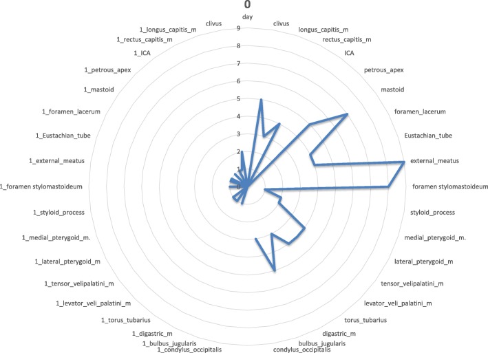
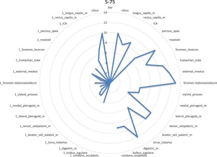
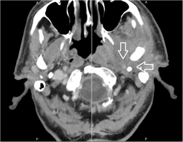
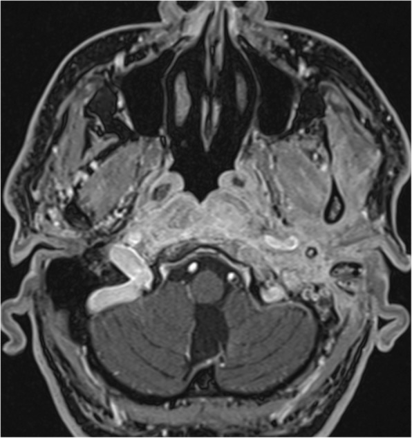
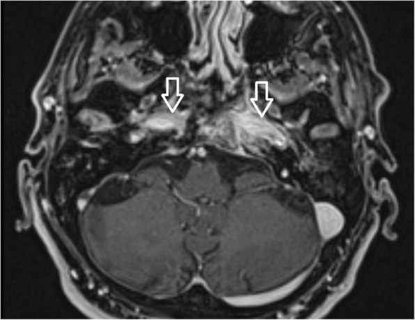
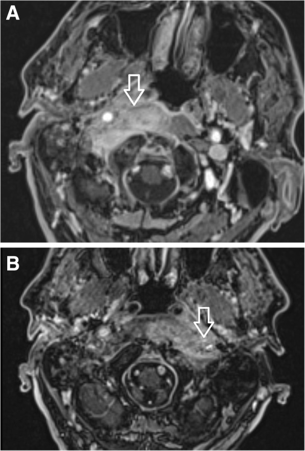
Results: In early stages of the disease, CT scan detects inflammatory changes closely related to the stylomastoid foramen and medially to the posterior belly of the digastric muscle, changes which have been proved to be crucial for the diagnosis of CSBO. Later the infection spreads to the contralateral side causing demineralisation of the bones.

Conclusion: Imaging methods play a crucial role not only in establishing the diagnosis, but also in anticipating the direction of infection spread underneath the skull base.

Keywords: Central skull-base osteomyelitis; Cranial nerve palsy, computed tomography; Imaging; Treatment.


Conflict of interest statement

All authors certify that they have no affiliations with or involvement in any organization or entity with any financial interest (such as honoraria; educational grants; participation in speakers’ bureaus; membership, employment, consultancies, stock ownership, or other equity interest; and expert testimony or patent-licensing arrangements), or non-financial interest (such as personal or professional relationships, affiliations, knowledge or beliefs) in the subject matter or materials discussed in this manuscript.

Figures

Fig. 1
Fig. 1
Day 0. Non-contrast enhanced CT in axial plane, section thickness 1 mm. The fatty tissues adjacent to the stylomastoid foramen are infiltrated, and cortical bone within the mastoid process is discretely eroded. The mastoid cells are filled with oedematous mucosa and soft tissue
Fig. 2
Fig. 2
Day 0 pattern of affected regions on the right side and on the contralateral left side. The outer ear canal and stylomastoid foramen are significantly affected
Fig. 3
Fig. 3
Day 5–75. Gadolinium-enhanced MRI, axial plane, T1-weighted, with fat suppression. Signal is homogeneously increased in the Eustachian tube and longus colli muscle. Cortical bone within the skull base is eroded, specifically the basal portion of the occipital bone ventrodorsally
Fig. 4
Fig. 4
Day 5–75: the inflammation spreads to further regions on the affected side
Fig. 5
Fig. 5
Contrast-enhanced axial CT image of temporal and occipital bones. Oedematous changes in soft tissues adjacent to the stylomastoid foramen on the left side represent the first sign of osteomyelitis spread beyond the temporal bone (arrows)
Fig. 6
Fig. 6
Day 93–177. Gadolinium-enhanced MRI, axial plane, T1-weighted, with fat suppression. The signal increases homogeneously on the left side in retrostyloid and pre-styloid regions, longus colli muscle, and Eustachian tubes on both sides. The signal mildly and homogeneously increases in the temporomandibular joint, lateral pterygoid, and masseter muscles on the left side. Bone marrow shows signs of infiltration in the basal part of the occipital bone
Fig. 7
Fig. 7
Day 93–177: massive spread of infection to the contralateral side (to the left)
Fig. 8
Fig. 8
Gadolinium-enhanced axial T1-weighted spin-echo image with fat saturation. In advanced stages of an infection, a butterfly-shaped soft tissue enhancement was described underneath both petrosal bones and the clivus (arrow)
Fig. 9
Fig. 9
Day 209–477. Gadolinium-enhanced MRI, axial plane, T1-weighted, with fat suppression. Mild homogeneous signal increase persists in the pharyngobasilar fascia and longus colli muscles on both sides. A cyst in the right maxillary sinus represents a secondary finding
Fig. 10
Fig. 10
Day 209–477: slow infection improvement in all regions on affected and contralateral sides
Fig. 11
Fig. 11
Patient No: 3, five months following mastoidectomy and antibiotic treatment: amoxicillin/clavulanic acid, gentamycin, clarithromycin and dalacin. Non-enhanced CT image (bone window) axial scan cross section of temporal and occipital bones. Osteolysis of the compact bone in the left lateral skull base represents a late sign of osteomyelitis (arrow). Minor changes of spongy bone and major changes of compact bone highlight spread of infection under the periosteum, but represents a late sign of the infection
Fig. 12
Fig. 12
a Patient 1, 2 months after the first clinical symptoms: MRI of the skull base. Contrast-enhanced axial T1-weighted spin-echo image with fat saturation. Inflammatory changes of the right side soft tissues beneath the skull base, musculus longus capitis and musculus rectus capitis anterior (arrow). The swelling extends to the midline; the infection originates from the right external auditory canal. b Patient No. 1, 4 months following the first clinical symptoms: MRI of the skull base of the same patient. Contrast-enhanced axial T1-weighted spin-echo image with fat saturation. After antibiotic treatment: ceftazidime, ciprofloxacin, clindamycin, Oxacyllin. Inflammatory changes of the soft tissues below the skull base progressed to the left side (arrow), musculus longus capitis and musculus rectus capitis anterior. The swelling affects the retropharyngeal and retrostyloid part of the parapharyngeal space in the midline and progresses to the left side

References

    1. Ridder GJ, Breunig C, Kaminsky J, Pfeiffer J. Central skull base osteomyelitis: new insights and implications for diagnosis and treatment. Eur Arch Otorhinolaryngol. 2015;272(5):1269–1276. doi: 10.1007/s00405-014-3390-y. - DOI - PubMed
    1. Johnson AK, Batra PS. Central skull base osteomyelitis: an emerging clinical entity. Laryngoscope. 2014;124(5):1083–1087. doi: 10.1002/lary.24440. - DOI - PubMed
    1. Gabrielli E, Fothergill AW, Brescini L, Sutton DA, Marchionni E, Orsetti E, Staffolani S, Castelli P, Gesuita R, Barchiesi F. Osteomyelitis caused by Aspergillus species: a review of 310 reported cases. Clin Microbiol Infect. 2014;20(6):559–565. doi: 10.1111/1469-0691.12389. - DOI - PubMed
    1. Razek A, Ghonim MR, Ashraf B. Computed tomography staging of middle ear cholesteatoma. Pol J Radiol. 2015;80:328–333. doi: 10.12659/PJR.894155. - DOI - PMC - PubMed
    1. Razek A. Assessment of masses of the external ear with diffusion-weighted MR imaging. Otol Neurotol. 2018;39(2):227–231. doi: 10.1097/MAO.0000000000001629. - DOI - PubMed

Publication types

MeSH terms