. 2019 Aug 1;10(1):231.
doi: 10.1186/s13287-019-1352-4.

Mesenchymal stromal cell-derived nanovesicles ameliorate bacterial outer membrane vesicle-induced sepsis via IL-10

Affiliations

Mesenchymal stromal cell-derived nanovesicles ameliorate bacterial outer membrane vesicle-induced sepsis via IL-10

Kyong-Su Park et al. Stem Cell Res Ther. .

Abstract

Background: Sepsis remains a source of high mortality in hospitalized patients despite proper antibiotic approaches. Encouragingly, mesenchymal stromal cells (MSCs) and their produced extracellular vesicles (EVs) have been shown to elicit anti-inflammatory effects in multiple inflammatory conditions including sepsis. However, EVs are generally released from mammalian cells in relatively low amounts, and high-yield isolation of EVs is still challenging due to a complicated procedure. To get over these limitations, vesicles very similar to EVs can be produced by serial extrusions of cells, after which they are called nanovesicles (NVs). We hypothesized that MSC-derived NVs can attenuate the cytokine storm induced by bacterial outer membrane vesicles (OMVs) in mice, and we aimed to elucidate the mechanism involved.

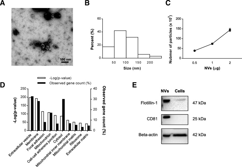
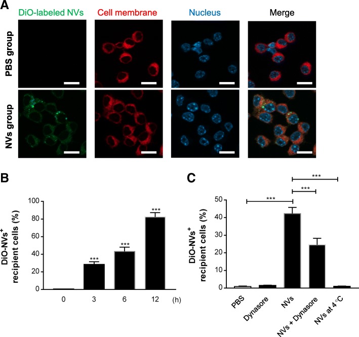
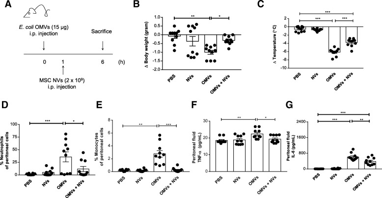
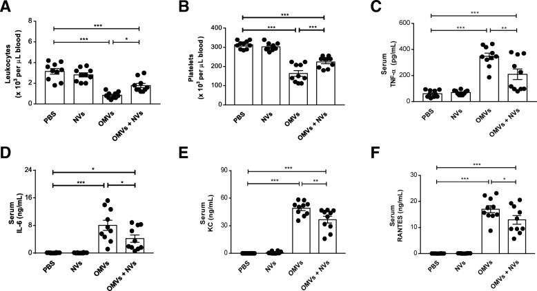
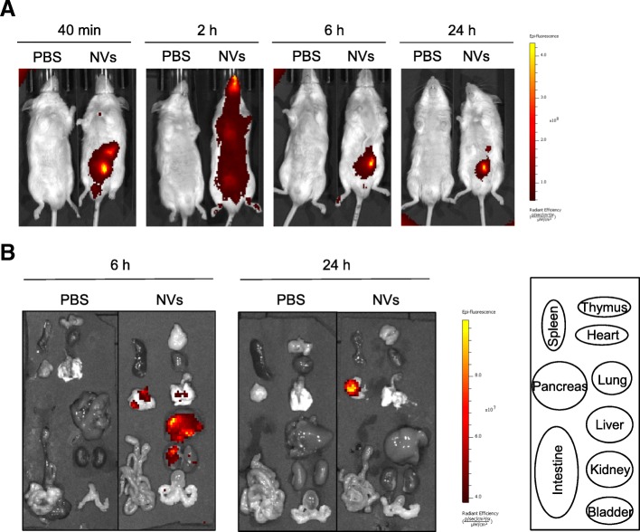
Methods: NVs were produced from MSCs by the breakdown of cells through serial extrusions and were subsequently floated in a density gradient. Morphology and the number of NVs were analyzed by transmission electron microscopy and nanoparticle tracking analysis. Mice were intraperitoneally injected with Escherichia coli-derived OMVs to establish sepsis, and then injected with 2 × 10⁹ NVs. Innate inflammation was assessed in peritoneal fluid and blood through investigation of infiltration of cells and cytokine production. The biodistribution of NVs labeled with Cy7 dye was analyzed using near-infrared imaging.

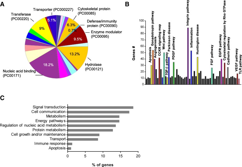
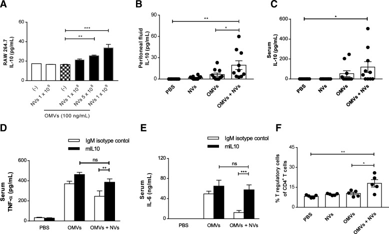
Results: Electron microscopy showed that NVs have a nanometer-size spherical shape and harbor classical EV marker proteins. In mice, NVs inhibited eye exudates and hypothermia, signs of a systemic cytokine storm, induced by intraperitoneal injection of OMVs. Moreover, NVs significantly suppressed cytokine release into the systemic circulation, as well as neutrophil and monocyte infiltration in the peritoneum. The protective effect of NVs was significantly reduced by prior treatment with anti-interleukin (IL)-10 monoclonal antibody. In biodistribution study, NVs spread to the whole mouse body and localized in the lung, liver, and kidney at 6 h.

Conclusions: Taken together, these data indicate that MSC-derived NVs have beneficial effects in a mouse model of sepsis by upregulating the IL-10 production, suggesting that artificial NVs may be novel EV-mimetics clinically applicable to septic patients.

Keywords: Anti-inflammation; Extracellular vesicles; Mesenchymal stromal cells; Nanovesicles; Outer membrane vesicles; Sepsis.


Conflict of interest statement

Jan Lötvall and Su Chul Jang have filed multiple patents for the development of EVs and EV-like nanovesicles for therapeutic purposes. Kyong-Su Park has filed several patents for using OMVs as sepsis model and vaccine, as well as investigating nanovesicle therapeutics. Jan Lötvall has previously been an employee of Codiak BioSciences Inc., and Su Chul Jang is currently employed by this company with the objective to develop EVs as therapeutics. The other authors declare that they have no competing interests.

Figures

Fig. 1
Fig. 1
Characterization of MSC-derived NVs. a TEM image of purified NVs. Scale bar, 100 nm. b Size distribution of NVs according to diameter. c The number of particles per proteins of NVs measured by nanoparticle tracking analysis (n = 3). d Top 10 subcellular localization GO terms enriched in NV proteome (based on P value). Three biological replicates were used, and commonly identified proteins in three runs were analyzed for GO terms. e Western blot analysis of EV markers on NV (10 μg) and MSC lysates (25 μg)
Fig. 2
Fig. 2
Functional analysis of NV proteome. a Distribution of the 2478 proteins sorted from all the NV proteins in different protein class by Panther analysis. b Panther pathway analysis of the NV proteome. c GO analysis of biological function of NV proteins conducted with Funrich software. The bar chart shows the top ten enriched categories (P < 0.05). Three biological replicates were used, and commonly identified proteins in three runs were analyzed for functional analysis
Fig. 3
Fig. 3
NVs are taken up by macrophage cells via endocytic pathway. a NVs (1 × 109) were incubated with RAW 264.7 cells for 6 h. NVs, cell membrane, and nuclei were stained by DiO (green), Cellmask Deep Red (red), and DAPI (blue), respectively. Scale bars, 20 μm. b RAW 264.7 cells were treated with DiO-labeled NVs for 0, 3, 6, and 12 h; then the uptake of the labeled NVs by cells was analyzed with flow cytometry; and data show the percentage of DiO-positive cells of three independent experiments. c Cells were pretreated with dynasore for 1 h at 37 °C, followed by incubation with DiO-labeled NVs for 6 h at 37 °C. Additionally, NVs were treated to the cells for 6 h at 4 °C. The uptake of the fluorescently labeled NVs by cells was analyzed with flow cytometry, and data represent the percentage of DiO-positive cells of three independent experiments. ***P < 0.001; versus the 0 h group. Error bars indicate SEM
Fig. 4
Fig. 4
Protective effect of NVs on peritoneal inflammation in OMV-induced sepsis in mice. a Study design for investigation of the therapeutic effect of NVs. Sublethal dose of OMVs (15 μg) from E. coli was injected i.p. once, followed by i.p. injection of NVs (2 × 109) at 1 h. Six hours after OMV injection, mice were sacrificed to check inflammatory parameters. b, c Body weight (b) and temperature (c) were measured at 6 h. d, e The percentage of neutrophils (d) and monocytes (e) in the peritoneum were determined by FACS analysis at 6 h following OMV administration. f, g Peritoneal fluid cytokines such as TNF-α (f) and IL-6 (g) were measured at 6 h. n = 10/group. *P < 0.05; **P < 0.01; ***P < 0.001. Error bars indicate SEM
Fig. 5
Fig. 5
NVs prevent the increase of systemic cytokines and chemokines provoked by OMVs. a, b The number of leukocytes (a) and platelets (b) was counted in blood at 6 h following OMV injection. cf The effect of NVs on the concentrations of TNF-α (c), IL-6 (d), KC (e), and RANTES (f) was analyzed in serum. n = 10/group. *P < 0.05; **P < 0.01; ***P < 0.001. Error bars indicate SEM
Fig. 6
Fig. 6
Biodistribution kinetic analysis of NVs in mice with near-infrared imaging. a The mice were injected by intraperitoneal injection of Cy7-labeled NVs (2 × 109) or PBS. At each time point, the fluorescence of the whole body was obtained by IVIS spectrum. b Various tissues were obtained at 6 and 24 h following injection of Cy7-labeled NVs. n = 3/group
Fig. 7
Fig. 7
IL-10-mediated immune suppression of NVs in OMV-induced sepsis. a RAW 264.7 cells were pre-incubated with OMVs (100 ng/mL) for 3 h and treated with various doses of NVs for 15 h, and the concentration of IL-10 in the conditioned media was measured. n = 3/group. b, c The concentration of IL-10 was evaluated in the peritoneal fluid (b) and serum (c) at 6 h following OMVs administration. n = 10/group. d, e mIL-10 or isotype-matched control antibody was intraperitoneally injected together with OMVs, followed by injection NVs after 1 h. Serum concentrations of TNF-α (d) and IL-6 (e) were measured at 6 h. n = 5/group. f The percentage of T regulatory cells in the peritoneum were determined by FACS analysis at 6 h. n = 5/group. *P < 0.05; **P < 0.01; ***P < 0.001; ns, not significant. Error bars indicate SEM

References

    1. Singer M, Deutschman CS, Seymour CW, Shankar-Hari M, Annane D, Bauer M, et al. The third international consensus definitions for sepsis and septic shock (Sepsis-3) JAMA. 2016;315:801–810. doi: 10.1001/jama.2016.0287. - DOI - PMC - PubMed
    1. Merx MW, Weber C. Sepsis and the heart. Circulation. 2007;116:793–802. doi: 10.1161/CIRCULATIONAHA.106.678359. - DOI - PubMed
    1. Nemeth K, Leelahavanichkul A, Yuen PS, Mayer B, Parmelee A, Doi K, et al. Bone marrow stromal cells attenuate sepsis via prostaglandin E (2)-dependent reprogramming of host macrophages to increase their interleukin-10 production. Nat Med. 2009;15:42–49. doi: 10.1038/nm.1905. - DOI - PMC - PubMed
    1. Hall SR, Tsoyi K, Ith B, Padera RF, Jr, Lederer JA, Wang Z, et al. Mesenchymal stromal cells improve survival during sepsis in the absence of heme oxygenase-1: the importance of neutrophils. Stem Cells. 2013;31:397–407. doi: 10.1002/stem.1270. - DOI - PMC - PubMed
    1. Lombardo E, van der Poll T, DelaRosa O, Dalemans W. Mesenchymal stem cells as a therapeutic tool to treat sepsis. World J Stem Cells. 2015;7:368–379. doi: 10.4252/wjsc.v7.i2.368. - DOI - PMC - PubMed

Publication types

MeSH terms