

Machine learning and glioma imaging biomarkers
- PMID: 31371027
- PMCID: PMC6927796
- DOI: 10.1016/j.crad.2019.07.001
Machine learning and glioma imaging biomarkers
Abstract
Aim: To review how machine learning (ML) is applied to imaging biomarkers in neuro-oncology, in particular for diagnosis, prognosis, and treatment response monitoring.
Materials and methods: The PubMed and MEDLINE databases were searched for articles published before September 2018 using relevant search terms. The search strategy focused on articles applying ML to high-grade glioma biomarkers for treatment response monitoring, prognosis, and prediction.
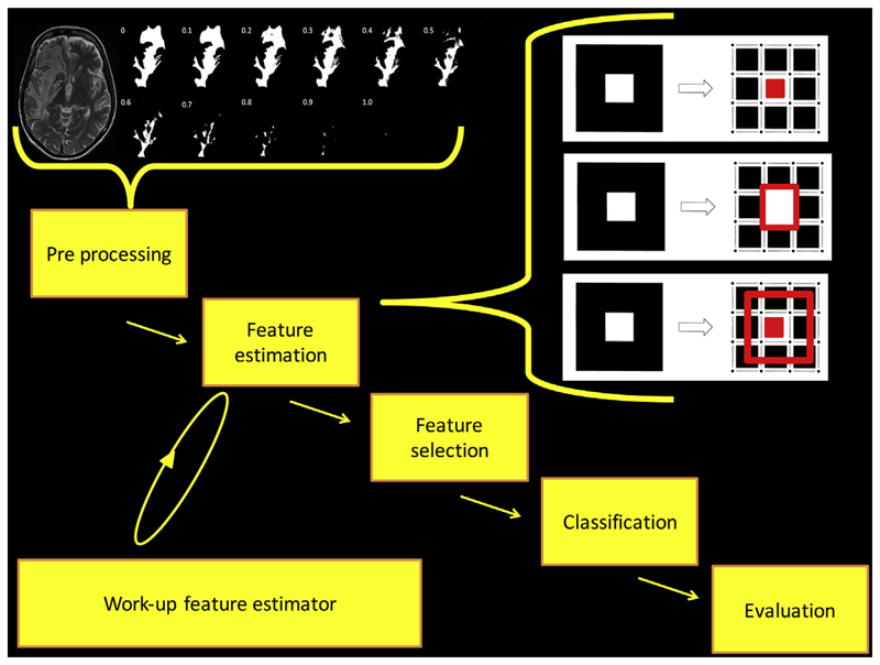
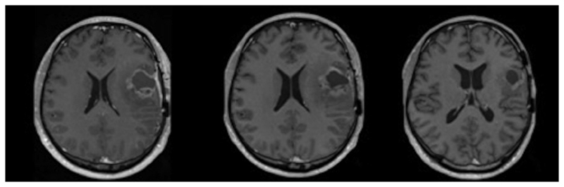
Results: Magnetic resonance imaging (MRI) is typically used throughout the patient pathway because routine structural imaging provides detailed anatomical and pathological information and advanced techniques provide additional physiological detail. Using carefully chosen image features, ML is frequently used to allow accurate classification in a variety of scenarios. Rather than being chosen by human selection, ML also enables image features to be identified by an algorithm. Much research is applied to determining molecular profiles, histological tumour grade, and prognosis using MRI images acquired at the time that patients first present with a brain tumour. Differentiating a treatment response from a post-treatment-related effect using imaging is clinically important and also an area of active study (described here in one of two Special Issue publications dedicated to the application of ML in glioma imaging).
Conclusion: Although pioneering, most of the evidence is of a low level, having been obtained retrospectively and in single centres. Studies applying ML to build neuro-oncology monitoring biomarker models have yet to show an overall advantage over those using traditional statistical methods. Development and validation of ML models applied to neuro-oncology require large, well-annotated datasets, and therefore multidisciplinary and multi-centre collaborations are necessary.
Copyright © 2019 The Authors. Published by Elsevier Ltd.. All rights reserved.
Conflict of interest statement
Jorge Cardoso is involved in machine learning enterprise and business.
Figures
References
-
- FDA-NIH Biomarker Working Group. BEST (biomarkers, EndpointS, and other tools) resource. 1st. Silver Spring, MD: Food and Drug Administration (US), co-published by Bethesda, MD National Institutes of Health (US); 2016. - PubMed
-
- Waldman AD, Jackson A, Price SJ, et al. Quantitative imaging biomarkers in neuro-oncology. Nat Rev Clin Oncol. 2009;6:445–54. - PubMed
-
- Darlix A, Zouaoui S, Rigau V, et al. Epidemiology for primary brain tumors: a nationwide population-based study. J Neurooncol. 2017;131:525–46. - PubMed
Publication types
MeSH terms
Substances
Grants and funding
LinkOut - more resources
Full Text Sources
Other Literature Sources
Medical
