. 2019 Aug 29;146(16):dev172668.
doi: 10.1242/dev.172668.

Laminin α2 controls mouse and human stem cell behaviour during midbrain dopaminergic neuron development

Affiliations

Laminin α2 controls mouse and human stem cell behaviour during midbrain dopaminergic neuron development

Maqsood Ahmed et al. Development. .

Abstract

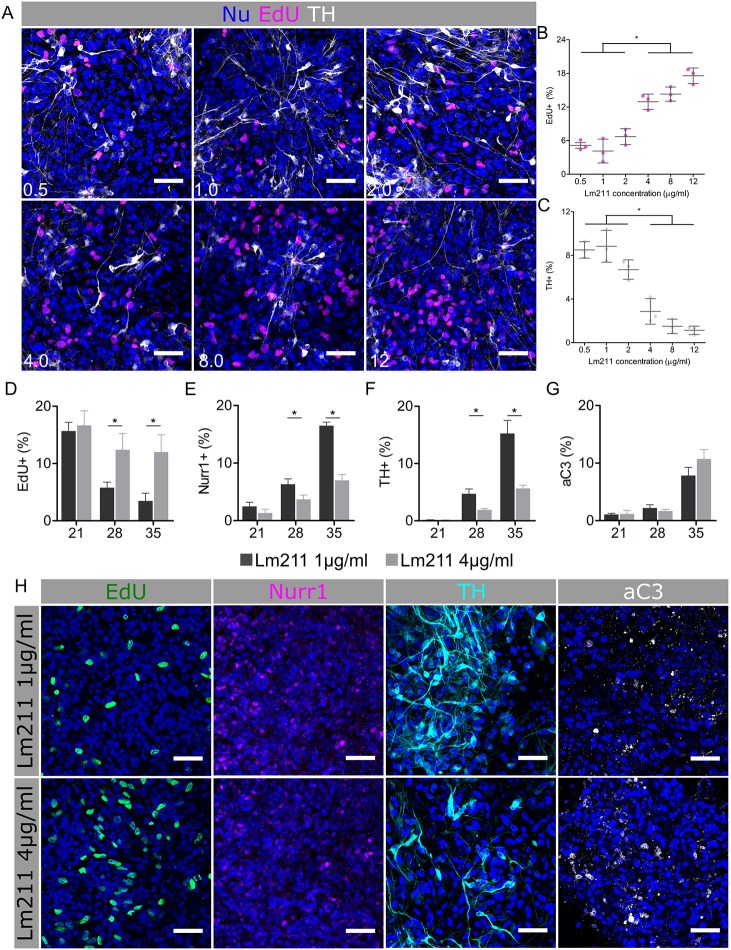
Development of the central nervous system requires coordination of the proliferation and differentiation of neural stem cells. Here, we show that laminin alpha 2 (lm-α2) is a component of the midbrain dopaminergic neuron (mDA) progenitor niche in the ventral midbrain (VM) and identify a concentration-dependent role for laminin α2β1γ1 (lm211) in regulating mDA progenitor proliferation and survival via a distinct set of receptors. At high concentrations, lm211-rich environments maintain mDA progenitors in a proliferative state via integrins α6β1 and α7β1, whereas low concentrations of lm211 support mDA lineage survival via dystroglycan receptors. We confirmed our findings in vivo, demonstrating that the VM was smaller in the absence of lm-α2, with increased apoptosis; furthermore, the progenitor pool was depleted through premature differentiation, resulting in fewer mDA neurons. Examination of mDA neuron subtype composition showed a reduction in later-born mDA neurons of the ventral tegmental area, which control a range of cognitive behaviours. Our results identify a novel role for laminin in neural development and provide a possible mechanism for autism-like behaviours and the brainstem hypoplasia seen in some individuals with mutations of LAMA2.

Keywords: Congenital muscular dystrophy; Dopaminergic neurons; Dystroglycan; Extracellular matrix; Integrin; Laminin; Neural stem cells.


Conflict of interest statement

Competing interestsThe authors declare no competing or financial interests.

Figures

Fig. 1.
Fig. 1.
Lm-α2 expression in human VM at 6 and 10 pcw. Lm-α2 expression (antibody, C13065) is enriched in human VM at 6 pcw, when it is composed of Ki67+ FoxA2+ progenitors. Expression diminishes by 10 pcw when the VM contains few Ki67+ cells and postmitotic Nurr1+ mDA neurons are established. scRNA-seq of human VM suggests that pericytes (hPeric) are the principal source of lm-α2. Right axis shows absolute molecule counts. Scale bars: 50 μm in all images.
Fig. 2.
Fig. 2.
Lm211 regulates proliferation and differentiation in a concentration-dependent manner. (A) Immunostaining of day 28 hESC-derived mDA cultures treated with lm211 over a concentration range of 0.5-12 μg ml−1. (B,C) Quantification of EdU (B) and TH (C) staining shows a significant increase in proliferating (EdU+) cells at lm211 concentrations of over 4 μg ml−1. The increase is associated with a reduction in TH+ mDA neurons (one-way ANOVA with Tukey’s post hoc test; P<0.0001, n=3). (D-G) Time series examining proliferation (D), differentiation (E,F) and survival (G) at days 21, 28 and 35 of culture at two different lm211 concentrations. mDA progenitors remain EdU+ at day 28 and 35 in the lm211-rich (4 μg ml−1) environment, resulting in significant reduction in the number of postmitotic mDA neuroblasts (Nurr1) and neurons (TH). There were no significant differences in aC3 staining between the two lm211 concentrations. Unpaired two-tailed t-test; *P<0.0001, n=3. (H) Representative images of day 35 cultures following lm211 treatment. Scale bars: 50 μm in all images.
Fig. 3.
Fig. 3.
Concentration-dependent effects of lm211 are mediated by distinct receptor engagement. (A) Immunostaining of day 28 cultures treated with lm211 (4 μg ml−1) in combination with integrin blocking antibodies and a control condition of lm211 (1 μg ml−1). (B,C) Quantification of proliferation (B) and differentiation (C) following blocking of integrin α-subunit demonstrates that blocking both integrins α6 and α7 reverses the effect of the lm211-rich environment. (D) Images of day 28 cultures treated with laminin isoforms (1 μg ml−1) and dystroglycan blocking antibody or isotype control. (E,F) Quantification of proliferation (E) and differentiation (F) demonstrates that blockade of dystroglycan reverses the gain in mDA neurons seen in response to treatment with lm211 compared with lm111 without effecting proliferation. Unpaired two-tailed t-test; *P<0.0001, n=3. Scale bars: 50 μm in all images.
Fig. 4.
Fig. 4.
Lm-α2 null VM is reduced in size with fewer cells and exhibits premature differentiation and depletion of the progenitor pool. Representative images and quantification of WT and mutant VMs at (A-D) E10.5 and (E-M) E12.5. (A) At E10.5, mutant embryos display a smaller ventral domain, as indicated by FoxA2 staining. (B-D) Quantification shows that fewer FoxA2+ cells are Ki67+ in the Lama2−/− embryo (B) but there are increased numbers of postmitotic mDA neuroblasts (Nurr1+; C) and mDA neurons (TH+; D). (E-J) By E12.5, there remain fewer proliferating FoxA2+ cells (F) and Lmx1a+ cells (G) in the VM of mutant embryos. (H-J) The intermediate and marginal zones of the mutant VM contain fewer Nurr1+ (H) and TH+ (I) cells, whereas there is an increase in apoptotic (aC3+) cells (J) at the ventricular zone and intermediate zone border in the absence of lm-α2. (K-M) Although there is a reduction in the number of Lmx1a+ mDA progenitors, the number of Lmx1a+ Ngn2+ cells remains the same (L), resulting in a greater proportion of progenitors undergoing neurogenesis [12.51±0.89% (WT) versus 20.73±3.49% (Lama2−/−)] in the mutant embryos. (M) EdU labelling confirmed this with a significant increase in the number of EdU+ Nurr1+ cells in the mutant embryos [512±41 (WT) versus 663±88 (Lama2−/−)], whereas there was a significant decrease in the number of Lmx1a+ EdU+ cells that are Nurr1 [201±35 (WT) versus 116±26 (Lama2−/−)]. Unpaired two-tailed t-test; *P<0.01, n=4-6. Scale bars: 50 μm in all images.
Fig. 5.
Fig. 5.
The midbrain of lm-α2 null brains contains fewer late-born calbindin+ mDA neurons of the VTA. (A-E) Immunostaining of postnatal brains (P15) of WT and mutant mice (A) shows fewer TH+ mDA neurons in Lama2−/− brains (B). In quantifying mDA subtype, there is a modest reduction in the number of Girk2+ mDA neurons (C) but a dramatic loss in calbindin+ mDA neurons (D) located medially in the VM. In normalising for the number of mDA neurons (E), there is a significant increase (P=0.0033) in the proportion of Girk2+ TH+ mDA neurons in the mutant mice concomitant with a reduction (P=0.0105) in calbindin+ TH+ mDA neurons. Unpaired two-tailed t-test; n=3 (WT), n=4 (KO). Scale bars: 100 μm in all images unless stated.

References

    1. Ahmed M. and ffrench-Constant C. (2016). Extracellular matrix regulation of stem cell behavior. Curr Stem Cell Rep. 2, 197-206. 10.1007/s40778-016-0056-2 - DOI - PMC - PubMed
    1. Alexander G. E. (2004). Biology of Parkinson's disease: pathogenesis and pathophysiology of a multisystem neurodegenerative disorder. Dialogues Clin. Neurosci. 6, 259-280. - PMC - PubMed
    1. Anderson C., Winder S. J. and Borycki A.-G. (2007). Dystroglycan protein distribution coincides with basement membranes and muscle differentiation during mouse embryogenesis. Dev. Dyn. Off. Publ. Am. Assoc. Anat. 236, 2627-2635. 10.1002/dvdy.21259 - DOI - PubMed
    1. Bayer S. A., Wills K. V., Triarhou L. C. and Ghetti B. (1995). Time of neuron origin and gradients of neurogenesis in midbrain dopaminergic neurons in the mouse. Exp. Brain Res. 105, 191-199. 10.1007/BF00240955 - DOI - PubMed
    1. Bedford L., Walker R., Kondo T., Van Cruchten I., King E. R. and Sablitzky F. (2005). Id4 is required for the correct timing of neural differentiation. Dev. Biol. 280, 386-395. 10.1016/j.ydbio.2005.02.001 - DOI - PubMed

Publication types

LinkOut - more resources