. 2019 Sep 2;218(9):3134-3152.
doi: 10.1083/jcb.201906059. Epub 2019 Aug 1.

Metalloprotease inhibitor TIMP proteins control FGF-2 bioavailability and regulate skeletal growth

Affiliations

Metalloprotease inhibitor TIMP proteins control FGF-2 bioavailability and regulate skeletal growth

Sanjay Saw et al. J Cell Biol. .

Abstract

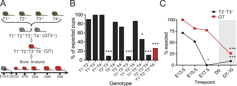
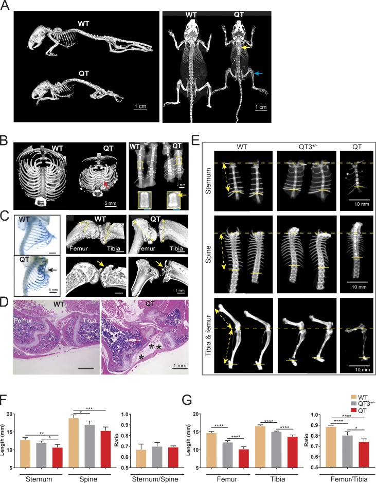
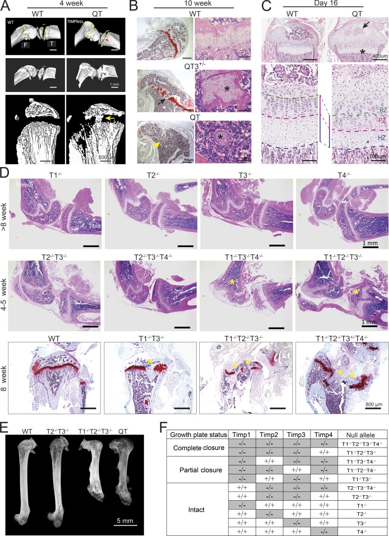
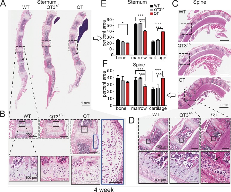
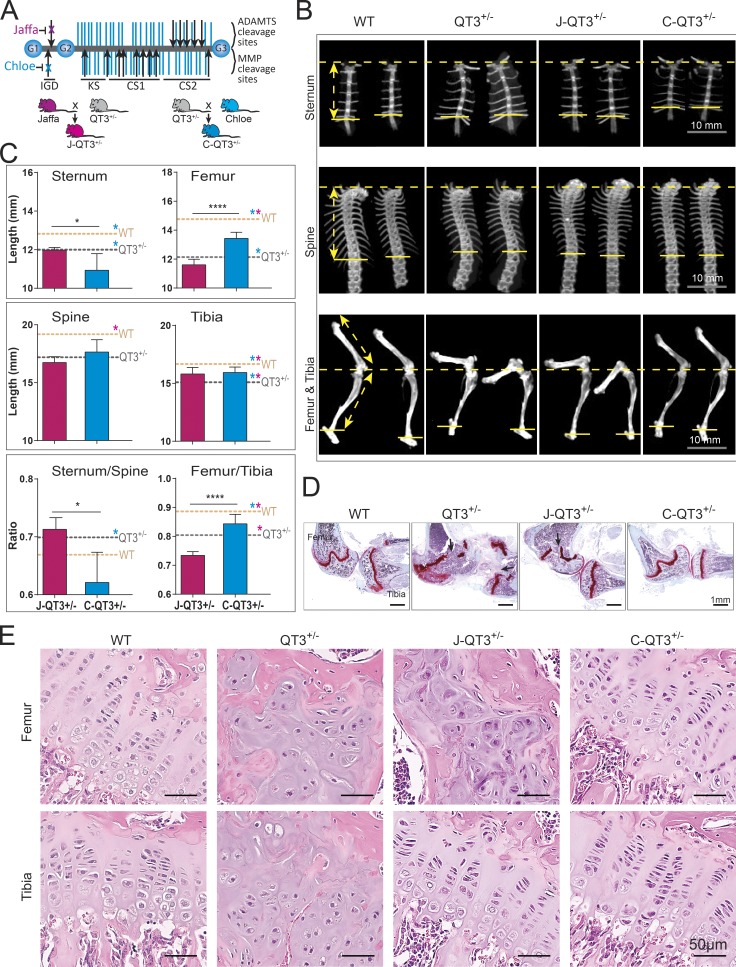
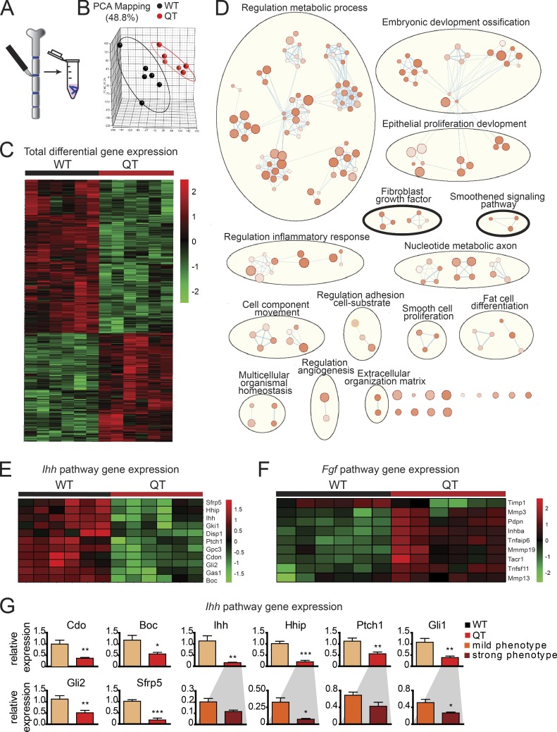
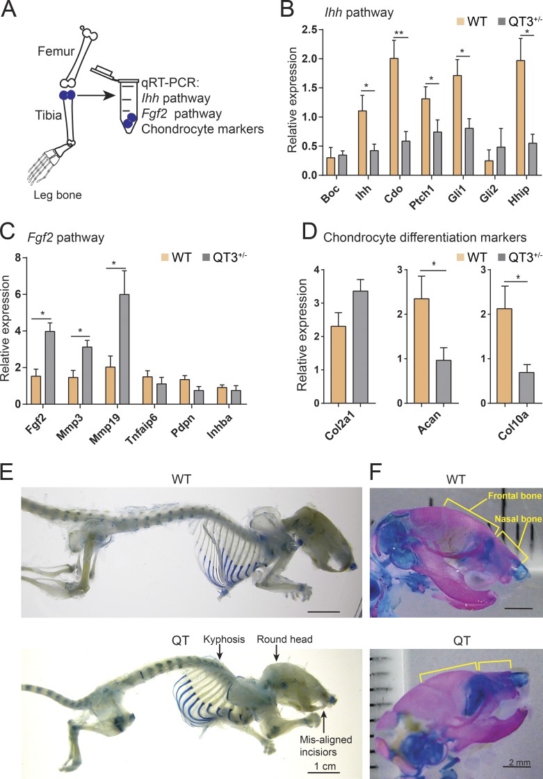
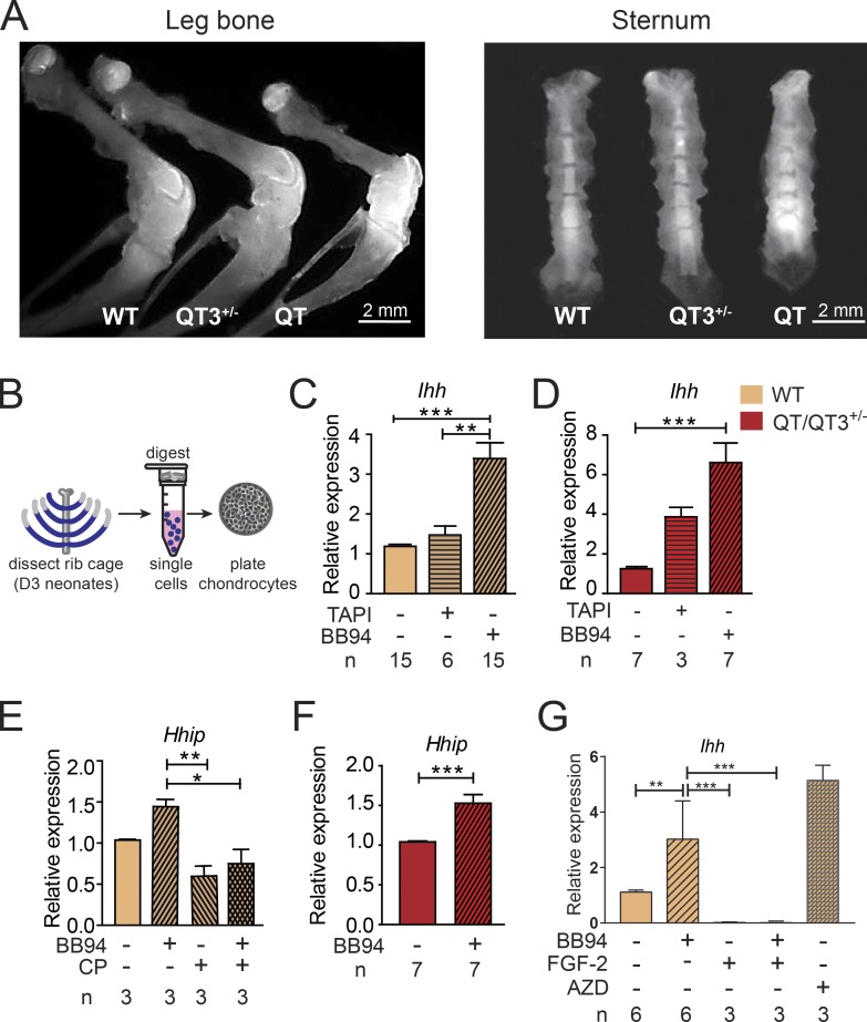
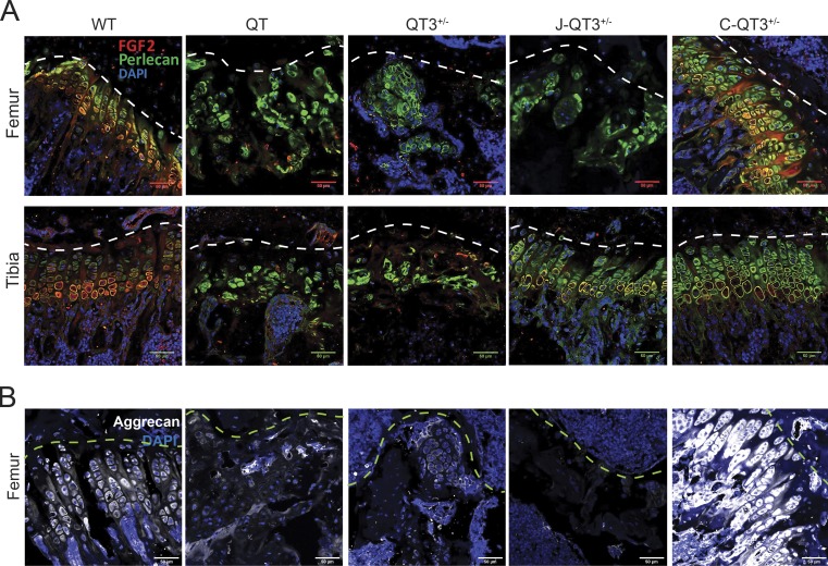
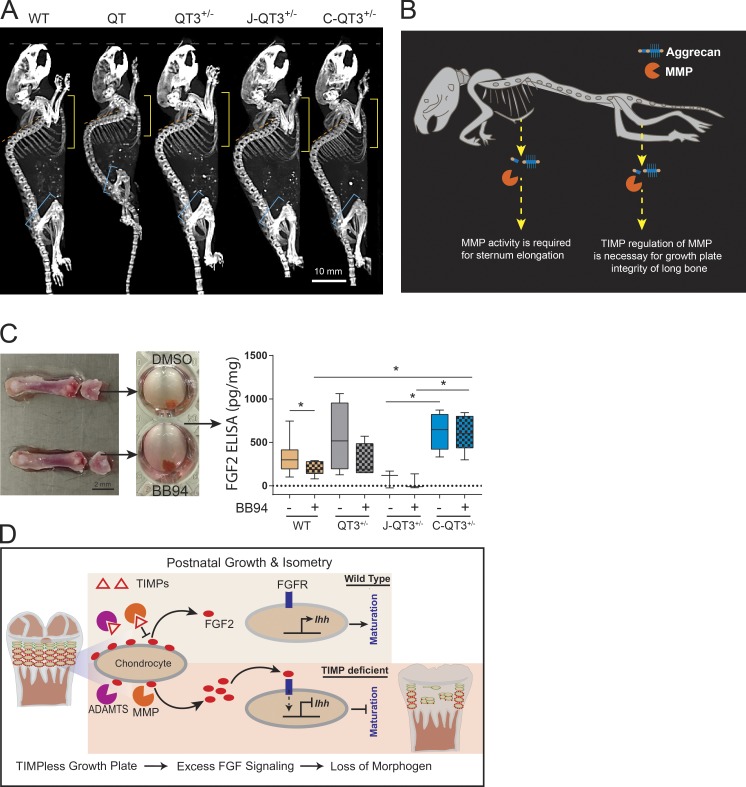
Regulated growth plate activity is essential for postnatal bone development and body stature, yet the systems regulating epiphyseal fusion are poorly understood. Here, we show that the tissue inhibitors of metalloprotease (TIMP) gene family is essential for normal bone growth after birth. Whole-body quadruple-knockout mice lacking all four TIMPs have growth plate closure in long bones, precipitating limb shortening, epiphyseal distortion, and widespread chondrodysplasia. We identify TIMP/FGF-2/IHH as a novel nexus underlying bone lengthening where TIMPs negatively regulate the release of FGF-2 from chondrocytes to allow IHH expression. Using a knock-in approach that combines MMP-resistant or ADAMTS-resistant aggrecans with TIMP deficiency, we uncouple growth plate activity in axial and appendicular bones. Thus, natural metalloprotease inhibitors are crucial regulators of chondrocyte maturation program, growth plate integrity, and skeletal proportionality. Furthermore, individual and combinatorial TIMP-deficient mice demonstrate the redundancy of metalloprotease inhibitor function in embryonic and postnatal development.


Figures

Figure 1.
Figure 1.
TIMP redundancy for in utero survivability. (A) Breeding strategy for the generation of QT3+/− and QT mice. Individual TIMP knockout mice were bred through several crosses to generate QT3+/− mice. QT3+/− were used as breeders to generate QT and QT3+/− mice. (B) Combinations of compound TIMP knockout mice were bred, and pups were genotyped. Number of pups genotyped at 1 wk after birth: T1−/− T2−/−, n = 60; T1−/− T3−/−, n = 45; T1−/− T4−/−, n = 103; T2−/− T3−/−, n = 590; T2−/− T4−/−, n = 80; T3−/− T4−/−, n = 85; T1−/− T2−/− T3−/−, n = 252; T1−/− T2−/− T4−/−, n = 14; T1−/− T3−/− T4−/−, n = 58; T2−/− T3−/− T4−/−, n = 137; T1−/− T2−/− T3−/− T4−/−, n = 473. χ2 test compared observed versus expected ratios for all genotypes. Comparison of expected (Mendelian distribution) versus observed revealed that the T2−/−T3−/− combination results in near-complete in utero lethality (9 observed of 98 expected from 590 born pups). Other non-Mendelian genotypes were T1−/− T2−/− T3−/− (3 of 37 from 252 born), T2−/− T3−/− T4−/− (3 of 26 from 137 born), and T1−/− T2−/− T3−/− T4−/− (31 of 118 from 473 born). (C) Embryos were genotyped and observed versus expected ratios compared using the χ2 test. Gray shading distinguishes postnatal time points. Enumeration of embryos (E13.5, E15.7, and E17.5) and born offspring showed that most T2−/− T3−/− die by E17.5, in contrast to QT, which were observed at >75% of the expected number. Therefore, the loss of TIMP2 and TIMP3 is detrimental at late gestation, while further additive loss of both TIMP1 and TIMP4 rescues lethality. It is conceivable that networks causing lethality in the T2−/− T3−/− scenario are either bypassed or opposed by the new milieu generated by complete TIMP loss. Numbers examined at E13.5, T2−/− T3−/− (n = 17) and T1−/− T2−/− T3−/− T4−/− (n = 13); E15.5, T2−/− T3−/− (n = 31) and T1−/− T2−/− T3−/− T4−/− (n = 81); E17.5, T2−/− T3−/− (n = 43), T1−/− T2−/− T3−/− T4−/− (n = 58); postnatal, T2−/− T3−/− (n = 33) and T1−/− T2−/− T3−/− T4−/− (n = 473). *, P < 0.05; ***, P < 0.001.
Figure 2.
Figure 2.
TIMPs are required for postnatal growth and long bone isometry. (A) Micro-CT images of WT and QT mice skeleton (age 8 wk) depict the short stature of QT mice as well as altered bone density (yellow arrow, brighter bone indicates higher density; blue arrow, lower density) in axial and appendicular bones. (B) Micro-CT scan of the thoracic girdle (left panel), presented after 3D isosurface rendering (4-wk-old WT and QT mice; transverse view). Red arrow points to a bony enlargement of rib-heads in QT mice. Faxitron x-ray images of 4-wk-old sternums (2 WT and 2 QT; right panel). The bright bands seen in enlargement reflect calcification at the edges of QT sternebrae (yellow arrow). (C) Alcian blue–stained thoracic girdles of 7-wk-old mice display Pectus formation in sternum of QT mice (black arrow; left panel). Micro-CT images of knee joints of 7-wk-old WT and QT mice (right panel). QT mice show metaphyseal flaring (double-sided yellow arrow) in femur (distal) and tibia (proximal) head. Yellow arrow indicates epiphysis of WT and loss of epiphysis in QT knee bone. (D) H&E-stained images of knee joints of 7-wk-old WT and QT mice display structural abnormality of QT knee joint. QT joint shows altered epiphysis (*), accompanied by fibrosis (4×/0.5-NA objective). (E) Micro-CT images of representative sternums (top), spines (middle), and leg bones (lower) of 8-wk-old WT, QT3+/−, and QT mice, depicting shortening of bone in TIMP-deficient mice. The distance from the top of sternum to the last attached rib was used to determine the length of sternum in different genotypes (double-sided yellow arrow). Similarly, the distance from the first attached rib to the last attached rib was measured to determine thoracic spine length for comparative purposes (double-sided yellow arrow). Leg bone length measurement depicted by double-headed yellow arrow. (F) Axial bone (sternum and thoracic spine) length and their proportionality in 8-wk-old WT (n = 7), QT3+/− (n = 5), and QT (n = 3) mice. Mean values of each dataset are plotted in graphs with error bars representing SEM. Datasets were compared by one-way ANOVA following Sidak’s multiple comparison test, *, P < 0.05; **, P < 0.01; ***, P < 0.001; ****, P < 0.0001. (G) Leg bone (femur and tibia) length and their proportionality in 8-wk-old WT (n = 7), QT3+/− (n = 5), and QT (n = 3) mice. Mean values of each dataset are plotted in graphs with error bars representing SEM. Datasets were compared by one-way ANOVA following Sidak’s multiple comparison test: *, P < 0.05; **, P < 0.01; ***, P < 0.001; ****, P < 0.0001.
Figure 3.
Figure 3.
Growth plate closure in different combination of TIMP knockouts. (A) Micro-CT images of 4-wk-old WT and QT knee joint. QT mice exhibit growth plate closure in the tibia and femur (upper and middle panels; cross section view; red arrow). High-resolution micro-CT images demonstrating growth plate closure in QT tibiae (yellow arrow; lower panel). (B) Femurs stained with H&E (right panel) and safranin-O (left panel) from 10-wk-old mice. WT mice retain an intact growth plate; QT3+/− cartilage is bridged by bone in several locations (black arrow), and only small islands of bone-encased cartilage remain in QT3+/− and QT bone (yellow arrow), as shown at higher magnification (*) on right panel. (C) H&E-stained tibiae of 16-d WT and QT mice. Upper panels indicate impaired secondary ossification center (arrow) and flattened growth plate (*) in QT tibia. Enlarged view in lower panels indicates that QT tibia has thinner hypertrophic zone (HZ) and proliferating zone (PZ). RZ, resting zone. (D) H&E-stained knee joints of different TIMP knockout combinations. Upper panels display knee joints of individual TIMP knockouts. Middle panels depict different levels of growth plate closure (*) of multiple-TIMP knockouts. Safranin O–stained tibiae of 8-wk-old WT and multiple TIMP knockouts (lower panels) exhibit growth plate closure (*; 4×/0.5-NA objective). (E) Faxitron x-ray image of femur, demonstrating the lengths of WT and different multiple TIMP knockouts (8-wk-old). (F) Summary of TIMP knockout combinations that result in growth plate closure or have an intact growth plate.
Figure 4.
Figure 4.
TIMPs are required for normal morphology of axial bone. (A–D) H&E-stained sagittal plane section of sternum (A and B) and spine (C and D; cervical and thoracic vertebrae) from 4-wk old mice. (B and D) Marked areas compare a single cartilaginous joint bordered by two growth plates. Further magnification exhibits cellular organization in growth plate cartilage and laterally migrated cartilage of QT sternum. Black arrow indicates loss of trabecular bone. (E and F) Histomorphometric quantification of percentage of cartilage, bone, and marrow in sternums and spine (7–8 wk-old; n = 3–6/group). ANOVA with Bonferroni’s multiple comparison test assessed significance for each tissue. *, P < 0.05; ***, P < 0.001.
Figure 5.
Figure 5.
Metalloprotease-resistant aggrecan rescues long bone proportionality. (A) Structure of aggrecan and the Chloe/Jaffa knock-in mutation. The aggrecan protein core (gray) contains three globular regions (G1–G3) and has ∼100 glycosaminoglycan chains (purple) attached. MMP and ADAMTS cleavage sites are indicated by black arrows. Disruption of specific cleavage sites by knock-in mutations are indicated by an x. Schematic of breeding strategies to generate J-QT3+/− and C-QT3+/− mice. CS1, chondroitin sulfate domain 1; CS2, chondroitin sulfate domain 2; KS, keratan sulfate. (B) Representative micro-CT images used to measure the length of sternum, the spine, and the leg bones of 8-wk old WT, QT3+/−, C-QT3+/−, and J-QT3+/−. Sternum (upper panel), spine (middle panel), and leg bones (lower panel). Arrows indicate measurements as described in legend of Fig. 1 H. (C) Quantification of axial and appendicular bones length in 8-wk-old WT, QT3+/−, C-QT3+/−, and J-QT3+/−. The sternum, spine, femur, and tibia are all shorter in the QT3+/− mouse compared with WT controls (dashed lines: WT, orange; QT3+/−, gray). C-QT3+/− mice have shorter sternums. Sternum:thoracic spine ratio indicates the effect was more pronounced in sternum than spine. C-QT3+/− partially rescued the shortened QT3+/− femur length, but J-QT3+/− mice could not. The femur:tibia ratio reflects that C-QT3+/− mice partially restored the leg bone compared with QT3+/− (n = 5–7/each group). Mean values of each dataset are plotted in graphs with error bars representing SEM. Datasets were compared by unpaired t test, *, P < 0.05; ****, P < 0.0001. Dunnett’s test P < 0.05 when WT or QT3+/− compared with C-QT3+/− (blue*) or J-QT3+/− (pink*). (D) Safranin O–stained knee joints of WT, QT3+/−, C-QT3+/−, and J-QT3+/− mice at 8 wk of age. Black arrows show growth plate closure in femur and tibia of QT3+/− mice; femur growth plate is rescued in the C-QT3+/− but not in J-QT3+/−, while the tibia growth plate is rescued by both mutant aggrecan (4×/0.5-NA objective). (E) H&E-stained femur and tibia growth plates of WT, QT3+/−, C-QT3+/−, and J-QT3+/− mice (8-wk-old) chondrocyte disorganization in the growth plates of QT3+/− femur and tibia, J-QT3+/− femur but not in the C-QT3+/− mice. Scale bar = 50 µm; 20×/0.5-NA objective.
Figure 6.
Figure 6.
Ihh down-regulation and higher FGF-2 signaling in QT chondrocytes. (A) Schematic of sternal cartilage macrodissection and RNA isolation. (B) Principal component analysis of microarray data from cartilage displaying distinct groups by genotype. Each datapoint represents one mouse. (C) Heat map of normalized mRNA levels of the 625 significantly altered genes between WT and QT samples (n = 6 each) determined by linear modeling with an adjusted P-value threshold of 0.05 and absolute fold change ≥1.5. (D) Gene enrichment analysis depicts altered biological processes in QT cartilage compared with WT. This analysis indicates a change in the bone development program in QT mice involving the IHH (smoothened) and FGF (encircled with black thick line) pathways. Size of nodes indicates number of genes involved, and color intensity indicates the q value (darker color indicates lower q value; q value cutoff is <0.05). (E) Heatmap of normalized mRNA levels of the IHH signaling pathway components, which are altered in QT versus WT samples. (F) Heatmap of normalized mRNA levels of FGF-2 target genes, in WT and QT cartilage. (G) Quantitative PCR confirming IHH pathway down-regulation (n = 6/genotype). Expression of genes cdo, Boc, Ihh, Hhip, Ptch1, Gli1, Gli2, and Sfrp5. Expression of Ihh, Hhip, Ptch1, and Gli1 were further subdivided by the severity of QT sternal phenotype (n = 3/group). Mean values of each dataset are plotted in graphs with error bars representing SEM. Data were compared using unpaired t test: *, P < 0.05; **, P < 0.01.
Figure 7.
Figure 7.
Ihh down-regulation and higher FGF-2 signaling in long bone chondrocyte. (A) Schematic of proximal tibial head RNA isolation for gene expression analysis. (B) Analysis of IHH pathway genes (Boc, Ihh, Cdo, Ptch1, Gli1, Gli2, Hhip) RT-PCR expression in proximal head of tibia of 4-wk-old WT and QT3+/− mice (n = 5/group). Mean values of each dataset are plotted in graphs with error bars representing SEM. Data were compared using unpaired t test: *, P < 0.05; **, P < 0.01. (C) Expression (RT-PCR) of FGF-2 pathway genes (Fgf2, Mmp3, Mmp9, Tnfaip6, Pdpn, and Inhba) in proximal head of tibia of 4-wk-old WT and QT3+/− mice (n = 4 for WT; n = 5 for QT3+/−). Mean values of each dataset are plotted in graphs with error bars representing SEM. Data were compared using unpaired t test: *, P < 0.05. (D) Chondrocyte development marker gene (Col2a1, Acan, and Col10a1) expression in proximal head of tibia of 4-wk-old WT and QT3+/− mice (n = 5/group). Values represent mean ± SEM; *, P < 0.05; **, P < 0.01. Mean values of each dataset are plotted in graphs with error bars representing SEM. Data were compared using unpaired t test: *, P < 0.05. (E) Alcian blue-stained skeleton of 8–9-wk-old WT and QT mice depicting round head, kyphosis, and misaligned incisors in QT mice. Scale bar = 1 cm. (F) Alcian blue– and alizarin red–stained skull of 1-d-old WT and QT mice depicting shortening of nasal and frontal bone. Scale bar = 2 mm.
Figure 8.
Figure 8.
TIMP/FGF-2/IHH comprise a critical axis for the growth plate integrity. (A) Metalloprotease activity detected in 4-wk-old bone, following in vivo injection of MMPSense750 fluorescent beacon. Ex vivo, growth plates of leg bone (left panel) display enhanced metalloprotease activity with progressively higher signal in QT3+/− and QT than WT. QT3+/− and QT sternum (right panel) also have higher metalloprotease activity than WT. (B) Schematic of chondrocyte isolation and culture. (C–F) Expression of Ihh and Hhip in chondrocyte cultures determined by RT-PCR. (C and D) Ihh expression following DMSO, BB94 (10 µM), or TAPI (20 µM) treatment in WT (C) and QT/QT3+/− (D) cultured chondrocytes. (E and F) Hhip expression following DMSO, BB94, or cyclopamine (CP; 10 µM) treatment in WT (E) and QT/QT3+/− (F) cultured chondrocytes. All reagents with media changed every 48 h over 7 d (n indicates number of independent mice). Mean values of each dataset are plotted in graphs with error bars representing SEM. Data were compared using one-way ANOVA following Dunnett’s multiple comparison test (C and D), Bonferroni’s multiple comparison test (E), and unpaired t test (F): **, P < 0.01; ***, P < 0.001. (G) Ihh expression following DMSO, BB94 (10 µM), FGF-2 (10 ng/ml), FGF-2 + BB94 (10 ng/ml + 10 µM), or AZD4547 (FGFR inhibitor; 500 nM) treatment in WT chondrocyte culture. All reagents with media changed every 48 h over 7 d (n indicates number of independent mice). Mean values of each dataset are plotted in graphs with error bars representing SEM. Datasets were compared by one-way ANOVA following Sidak’s multiple comparison test: *, P < 0.05; **, P < 0.01; ***, P < 0.001.
Figure 9.
Figure 9.
Protease-resistant aggrecans and FGF-2 levels. (A) Immunostaining of FGF-2 and perlecan in the femur (top panel) and tibia (bottom panel) of 8-wk-old WT, QT, QT3+/−, J-QT3+/−, and C-QT3+/− mice. FGF-2 staining and perlecan organization are restored in growth plate of C-QT3+/− femur but not of J-QT3+/−, and in the tibia growth plate in both J-QT3+/− and C-QT3+/− mice. Scale bar = 50 µm; 20×/0.8-NA objective. (B) Immunofluorescence of aggrecan in the femur of 8-wk-old WT, QT, QT3+/−, J-QT3+/−, and C-QT3+/− mice. Scale bar = 50 µm; 20×/0.8-NA objective.
Figure 10.
Figure 10.
Protease-mediated FGF-2 release and body stature. (A) Whole-body micro-CT images of 8-wk-old WT, QT, QT3+/−, J-QT3+/−, and C-QT3+/− mice. Images allow comparison of disproportion in the femur and tibia lengths, sternum, and spine curvature among different genotypes (yellow line, length of sternum; orange line, extent of kyphosis; blue line, femur length). Scale bar = 10 mm. (B) Cartoon depicting the effect of aggrecan cleavage by MMPs on bone length. The lack of MMP regulation by TIMPs (excess MMP activity) results in growth plate aberrations in the long bones, while the lack of MMP processing has the profound effect of shortening the sternum. (C) Distal femoral head explant culture to determine FGF-2 level by ELISA on the femoral distal head culture supernatant of WT (n = 10), QT3+/− (n = 4), J-QT3+/− (n = 3), and C-QT3+/− (n = 5) mice. Reduction of FGF-2 release from contralateral femoral head by BB94 treatment, determined by ratio paired t test. Each box of box-and-whisker plot shows second to third quartile of datasets and line within the box shows median; *, P < 0.05. (D) Pictorial summary delineating the role of TIMPs in molecular processing to alter the bone length. Activation of the FGFR by excessive metalloprotease release of perlecan-bound FGF2 represses IHH-mediated chondrocyte maturation.

Similar articles

Cited by

References

    1. Aiken A., and Khokha R.. 2010. Unraveling metalloproteinase function in skeletal biology and disease using genetically altered mice. Biochim. Biophys. Acta. 1803:121–132. 10.1016/j.bbamcr.2009.07.002 - DOI - PubMed
    1. Ali S.A., and Alman B.. 2012. RNA extraction from human articular cartilage by chondrocyte isolation. Anal. Biochem. 429:39–41. 10.1016/j.ab.2012.06.028 - DOI - PubMed
    1. Bogin B., and Varela-Silva M.I.. 2010. Leg length, body proportion, and health: a review with a note on beauty. Int. J. Environ. Res. Public Health. 7:1047–1075. 10.3390/ijerph7031047 - DOI - PMC - PubMed
    1. Börjesson A.E., Windahl S.H., Karimian E., Eriksson E.E., Lagerquist M.K., Engdahl C., Antal M.C., Krust A., Chambon P., Sävendahl L., and Ohlsson C.. 2012. The role of estrogen receptor-α and its activation function-1 for growth plate closure in female mice. Am. J. Physiol. Endocrinol. Metab. 302:E1381–E1389. 10.1152/ajpendo.00646.2011 - DOI - PMC - PubMed
    1. Briscoe J., and Thérond P.P.. 2013. The mechanisms of Hedgehog signalling and its roles in development and disease. Nat. Rev. Mol. Cell Biol. 14:416–429. 10.1038/nrm3598 - DOI - PubMed

Publication types

MeSH terms

Substances

Grants and funding