

Prion protein deficiency impairs hematopoietic stem cell determination and sensitizes myeloid progenitors to irradiation
- PMID: 31371412
- PMCID: PMC7193476
- DOI: 10.3324/haematol.2018.205716
Prion protein deficiency impairs hematopoietic stem cell determination and sensitizes myeloid progenitors to irradiation
Abstract
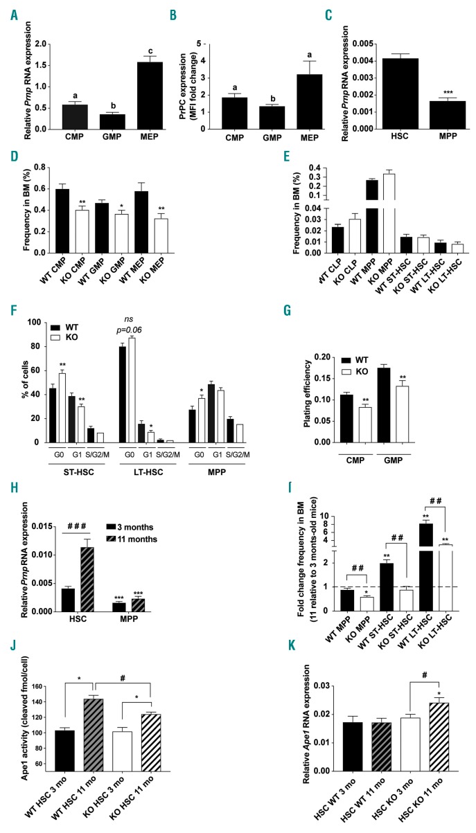
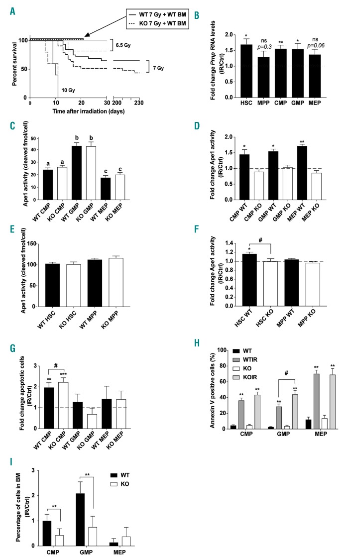
Highly conserved among species and expressed in various types of cells, numerous roles have been attributed to the cellular prion protein (PrPC). In hematopoiesis, PrPC regulates hematopoietic stem cell self-renewal but the mechanisms involved in this regulation are unknown. Here we show that PrPC regulates hematopoietic stem cell number during aging and their determination towards myeloid progenitors. Furthermore, PrPC protects myeloid progenitors against the cytotoxic effects of total body irradiation. This radioprotective effect was associated with increased cellular prion mRNA level and with stimulation of the DNA repair activity of the Apurinic/pyrimidinic endonuclease 1, a key enzyme of the base excision repair pathway. Altogether, these results show a previously unappreciated role of PrPC in adult hematopoiesis, and indicate that PrPC-mediated stimulation of BER activity might protect hematopoietic progenitors from the cytotoxic effects of total body irradiation.
Copyright© 2020 Ferrata Storti Foundation.
Figures
Similar articles
-
Prion protein expression in human leukocyte differentiation.Blood. 1998 Mar 1;91(5):1556-61. Blood. 1998. PMID: 9473220
-
An antipsychotic drug exerts anti-prion effects by altering the localization of the cellular prion protein.PLoS One. 2017 Aug 7;12(8):e0182589. doi: 10.1371/journal.pone.0182589. eCollection 2017. PLoS One. 2017. PMID: 28787011 Free PMC article.
-
Hhex Regulates Hematopoietic Stem Cell Self-Renewal and Stress Hematopoiesis via Repression of Cdkn2a.Stem Cells. 2017 Aug;35(8):1948-1957. doi: 10.1002/stem.2648. Epub 2017 Jun 19. Stem Cells. 2017. PMID: 28577303
-
Expression of cellular prion protein on blood cells: potential functions in cell physiology and pathophysiology of transmissible spongiform encephalopathy diseases.Transfus Med Rev. 2001 Oct;15(4):268-81. doi: 10.1053/tmrv.2001.26957. Transfus Med Rev. 2001. PMID: 11668434 Review.
-
Prion Protein in Glioblastoma Multiforme.Int J Mol Sci. 2019 Oct 15;20(20):5107. doi: 10.3390/ijms20205107. Int J Mol Sci. 2019. PMID: 31618844 Free PMC article. Review.
Cited by
-
Emerging Role of Cellular Prion Protein in the Maintenance and Expansion of Glioma Stem Cells.Cells. 2019 Nov 18;8(11):1458. doi: 10.3390/cells8111458. Cells. 2019. PMID: 31752162 Free PMC article. Review.
-
The multiple functions of PrPC in physiological, cancer, and neurodegenerative contexts.J Mol Med (Berl). 2022 Oct;100(10):1405-1425. doi: 10.1007/s00109-022-02245-9. Epub 2022 Sep 3. J Mol Med (Berl). 2022. PMID: 36056255 Review.
-
Prion infection modulates hematopoietic stem/progenitor cell fate through cell-autonomous and non-autonomous mechanisms.Leukemia. 2023 Apr;37(4):877-887. doi: 10.1038/s41375-023-01828-w. Epub 2023 Jan 27. Leukemia. 2023. PMID: 36707620 Free PMC article.
-
The Cellular Prion Protein and the Hallmarks of Cancer.Cancers (Basel). 2021 Oct 8;13(19):5032. doi: 10.3390/cancers13195032. Cancers (Basel). 2021. PMID: 34638517 Free PMC article. Review.
-
Do Transgenerational Epigenetic Inheritance and Immune System Development Share Common Epigenetic Processes?J Dev Biol. 2021 May 12;9(2):20. doi: 10.3390/jdb9020020. J Dev Biol. 2021. PMID: 34065783 Free PMC article. Review.
References
-
- Simonnet AJ, Nehmë J, Vaigot P, Barroca V, Leboulch P, Tronik-Le Roux D. Phenotypic and functional changes induced in hematopoietic stem/progenitor cells after gamma-ray radiation exposure. Stem Cells. 2009;27(6):1400–1409. - PubMed
-
- Down JD, Boudewijn A, van Os R, Thames HD, Ploemacher RE. Variations in radiation sensitivity and repair among different hematopoietic stem cell subsets following fractionated irradiation. Blood. 1995; 86(1):122–127. - PubMed
Publication types
MeSH terms
Substances
LinkOut - more resources
Full Text Sources
Molecular Biology Databases
