. 2019 Aug 20;116(34):16711-16716.
doi: 10.1073/pnas.1903500116. Epub 2019 Aug 1.

Building a synthetic mechanosensitive signaling pathway in compartmentalized artificial cells

Affiliations

Building a synthetic mechanosensitive signaling pathway in compartmentalized artificial cells

James W Hindley et al. Proc Natl Acad Sci U S A. .

Abstract

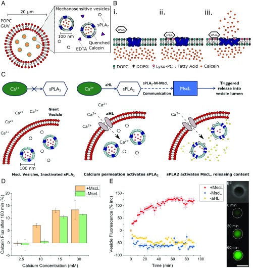
To date, reconstitution of one of the fundamental methods of cell communication, the signaling pathway, has been unaddressed in the bottom-up construction of artificial cells (ACs). Such developments are needed to increase the functionality and biomimicry of ACs, accelerating their translation and application in biotechnology. Here, we report the construction of a de novo synthetic signaling pathway in microscale nested vesicles. Vesicle-cell models respond to external calcium signals through activation of an intracellular interaction between phospholipase A2 and a mechanosensitive channel present in the internal membranes, triggering content mixing between compartments and controlling cell fluorescence. Emulsion-based approaches to AC construction are therefore shown to be ideal for the quick design and testing of new signaling networks and can readily include synthetic molecules difficult to introduce to biological cells. This work represents a foundation for the engineering of multicompartment-spanning designer pathways that can be utilized to control downstream events inside an AC, leading to the assembly of micromachines capable of sensing and responding to changes in their local environment.

Keywords: MscL; artificial cells; nested vesicle; phospholipase A2; signaling pathway.


Conflict of interest statement

The authors declare no conflict of interest.

Figures

Fig. 1.
Fig. 1.
Using the sPLA2–M–MscL network to build a synthetic mechanosensitive signaling pathway inside an artificial cell (AC). (A) Composition of the nested AC: A microscale POPC membrane encloses 1:1 DOPC:DOPG vesicles containing reconstituted mechanosensitive channel of large conductance (MscL), secretory phospholipase A2 (sPLA2) enzyme, and EDTA to chelate trace calcium present in the AC. (B) Function of the sPLA2–M–MscL network. (B, i) MscL is reconstituted into a DOPC:DOPG membrane and is closed in the absence of tension or asymmetry in the membrane. sPLA2 is added to the solution. (B, ii) sPLA2 binds to the membrane and begins to catalyze the production of LPC and a concomitant fatty acid. The asymmetric generation of LPC begins to asymmetrically change the pressure profile of the lipid bilayer. (B, iii) Once a critical amount of LPC has been produced, MscL responds to the lateral pressure change by opening to form a 3- to 4-nm diameter pore in the lipid bilayer, releasing encapsulated cargo across the membrane. (C) Proposed functioning of the synthetic mechanosensitive signaling pathway. Ca2+ is prevented from entering the nested vesicle due to the presence of the outer POPC membrane. Permeabilization of the outer membrane (here accomplished with αHL) then results in a calcium influx, activating latent sPLA2 in the vesicle lumen. This activates the sPLA2–M–MscL network, resulting in content release (and potentially the control of downstream events) within the AC. (D) Monitoring activation of the mechanosensitive pathway with fluorescence spectroscopy. Successful activation is triggered by the addition of ∼10 mM Ca2+. Error bars represent 1 SD (n = 3). (E) Confirming activation of the pathway through fluorescence microscopy of individual nested vesicles. (E, Left) Both MscL and αHL are necessary to increase vesicle fluorescence (red squares), while absence of MscL (blue circles) or αHL (yellow triangles) prevents network activation. Error bars represent 1 SEM (n = 15, 14, and 13, respectively). E, Right highlights micrographs of a nested vesicle in bright-field and fluorescence microscopy of pathway activation within the nested vesicle at t = 0, 30, and 60 min, respectively. (Scale bar, 10 μm.)
Fig. 2.
Fig. 2.
MscL is essential for sPLA2–M–MscL communication. (A) The release of calcein from 1:1 DOPC:DOPG vesicles can be monitored spectroscopically over time. sPLA2 is added at 10 min, before monitoring calcein fluorescence for 100 min. Release of calcein from vesicles results in a fluorescence increase as the dye dilutes in external solution and self-quenching becomes inefficient. Error bars show propagated error of 1 SD (n = 3). (B) Total calcein flux at 100 min for vesicles ± MscL. sPLA2-concentration-dependent calcein flux is only observed for vesicles containing MscL. Error bars show 1 SD (n = 3).
Fig. 3.
Fig. 3.
The sPLA2–M–MscL network can be controlled through calcium chelation. (A) sPLA2 inactivation, and hence network activation, can be achieved through the addition of increasing concentrations of the Ca2+ chelator EDTA. Error bars = 1 SD (n = 3). (B) Monitoring reactivation of the network using calcein flux after the addition of Ca2+ (0–10.0 mM) to solutions containing the network and 2.5 mM EDTA. Error bars show 1 SD (n = 3). (C) Total calcein flux after 60 min for 1:1 DOPC:DOPG vesicles ± MscL showing that the MscL channel is necessary for network reactivation with Ca2+. Error bars show 1 SD (n = 3).

Similar articles

Cited by

References

    1. Lai E. C., Notch signaling: Control of cell communication and cell fate. Development 131, 965–973 (2004). - PubMed
    1. Waters C. M., Bassler B. L., Quorum sensing: Cell-to-cell communication in bacteria. Annu. Rev. Cell Dev. Biol. 21, 319–346 (2005). - PubMed
    1. Ratajczak J., Wysoczynski M., Hayek F., Janowska-Wieczorek A., Ratajczak M. Z., Membrane-derived microvesicles: Important and underappreciated mediators of cell-to-cell communication. Leukemia 20, 1487–1495 (2006). - PubMed
    1. Rasmussen H., Cell communication, calcium ion, and cyclic adenosine monophosphate. Science 170, 404–412 (1970). - PubMed
    1. Clapham D. E., Calcium signaling. Cell 131, 1047–1058 (2007). - PubMed

Publication types

MeSH terms