

Retinal and optic nerve degeneration in liver X receptor β knockout mice
- PMID: 31371497
- PMCID: PMC6697819
- DOI: 10.1073/pnas.1904719116
Retinal and optic nerve degeneration in liver X receptor β knockout mice
Abstract
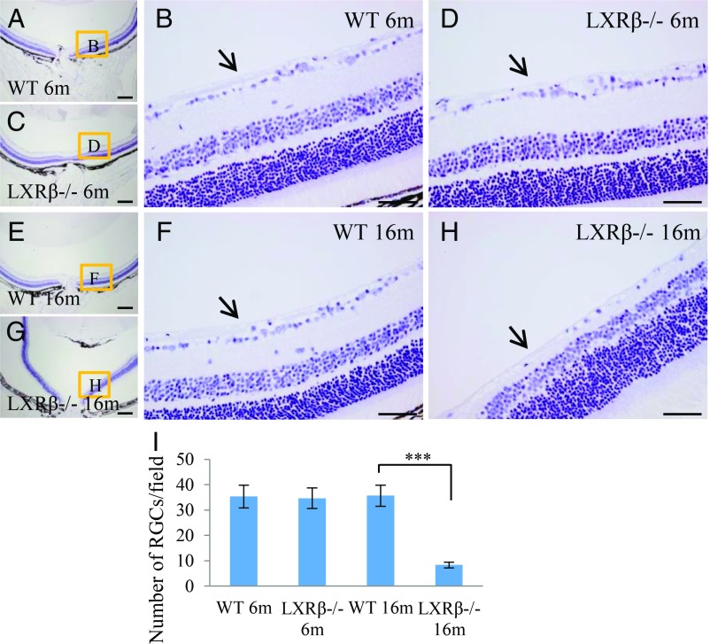
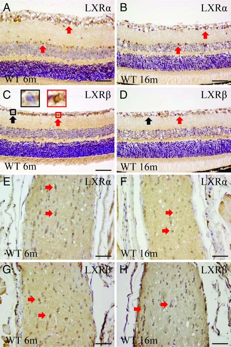
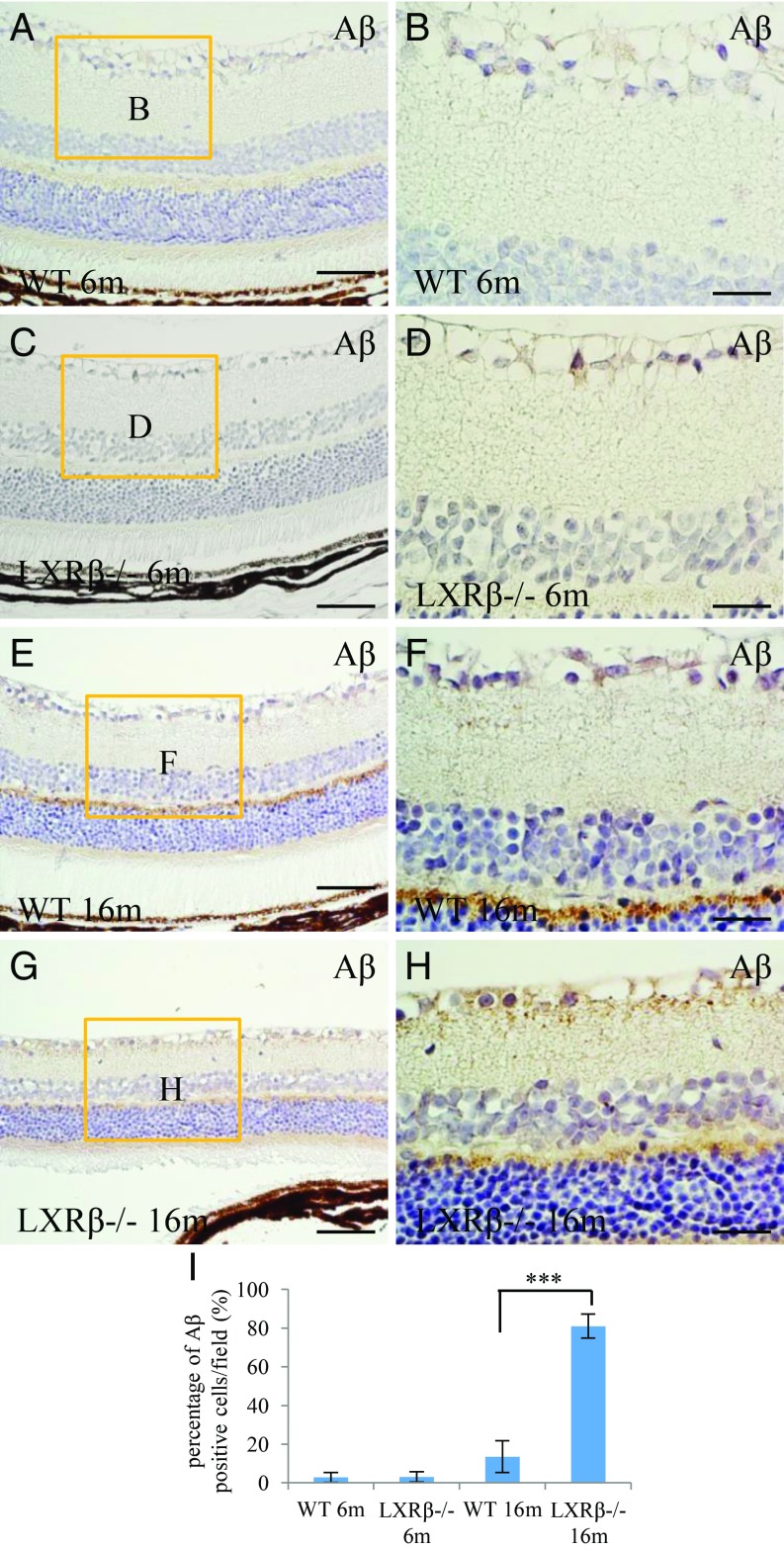
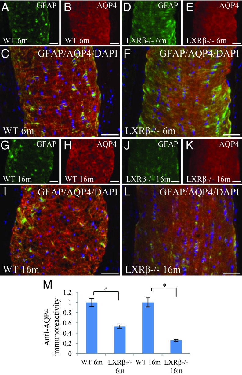
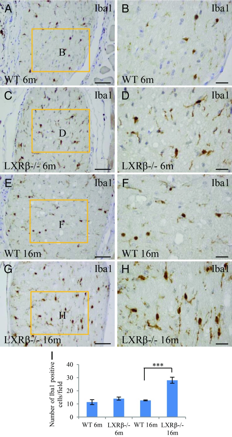
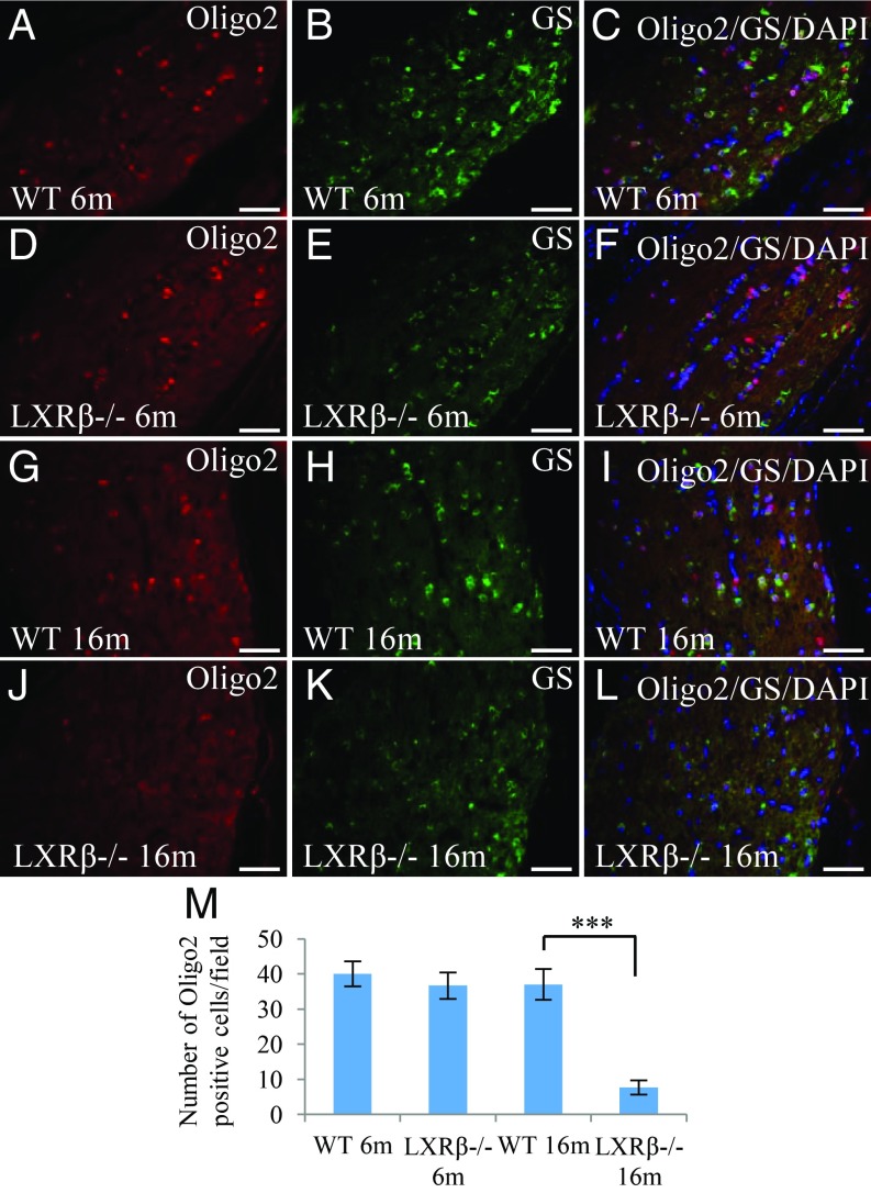
The retina is an extension of the brain. Like the brain, neurodegeneration of the retina occurs with age and is the cause of several retinal diseases including optic neuritis, macular degeneration, and glaucoma. Liver X receptors (LXRs) are expressed in the brain where they play a key role in maintenance of cerebrospinal fluid and the health of dopaminergic neurons. Herein, we report that LXRs are expressed in the retina and optic nerve and that loss of LXRβ, but not LXRα, leads to loss of ganglion cells in the retina. In the retina of LXRβ-/- mice, there is an increase in amyloid A4 and deposition of beta-amyloid (Aβ) aggregates but no change in the level of apoptosis or autophagy in the ganglion cells and no activation of microglia or astrocytes. However, in the optic nerve there is a loss of aquaporin 4 (AQP4) in astrocytes and an increase in activation of microglia. Since loss of AQP4 and microglial activation in the optic nerve precedes the loss of ganglion cells, and accumulation of Aβ in the retina, the cause of the neuronal loss appears to be optic nerve degeneration. In patients with optic neuritis there are frequently AQP4 autoantibodies which block the function of AQP4. LXRβ-/- mouse is another model of optic neuritis in which AQP4 antibodies are not detectable, but AQP4 function is lost because of reduction in its expression.
Keywords: aquaporin 4; nuclear receptor; optic neuritis; retinal degeneration; β amyloid.
Conflict of interest statement
The authors declare no conflict of interest.
Figures

Similar articles
-
Kidins220-deficient hydrocephalus mice exhibit altered glial phenotypes and AQP4 differential regulation in the retina and optic nerve, with preserved retinal ganglion cell survival.Fluids Barriers CNS. 2025 Feb 12;22(1):16. doi: 10.1186/s12987-025-00626-z. Fluids Barriers CNS. 2025. PMID: 39939990 Free PMC article.
-
BDNF impairment is associated with age-related changes in the inner retina and exacerbates experimental glaucoma.Biochim Biophys Acta. 2014 Sep;1842(9):1567-78. doi: 10.1016/j.bbadis.2014.05.026. Epub 2014 Jun 2. Biochim Biophys Acta. 2014. PMID: 24942931
-
The role of aquaporin-4 in optic nerve head astrocytes in experimental glaucoma.PLoS One. 2021 Feb 2;16(2):e0244123. doi: 10.1371/journal.pone.0244123. eCollection 2021. PLoS One. 2021. PMID: 33529207 Free PMC article.
-
Neuroinflammation in Glaucoma and Optic Nerve Damage.Prog Mol Biol Transl Sci. 2015;134:343-63. doi: 10.1016/bs.pmbts.2015.06.010. Epub 2015 Jul 10. Prog Mol Biol Transl Sci. 2015. PMID: 26310164 Review.
-
The role of autophagy in axonal degeneration of the optic nerve.Exp Eye Res. 2016 Mar;144:81-9. doi: 10.1016/j.exer.2015.08.016. Epub 2015 Aug 24. Exp Eye Res. 2016. PMID: 26315785 Review.
Cited by
-
Liver X Receptor Regulation of Glial Cell Functions in the CNS.Biomedicines. 2022 Sep 2;10(9):2165. doi: 10.3390/biomedicines10092165. Biomedicines. 2022. PMID: 36140266 Free PMC article. Review.
-
Loss of ERβ in Aging LXRαβ Knockout Mice Leads to Colitis.Int J Mol Sci. 2023 Aug 5;24(15):12461. doi: 10.3390/ijms241512461. Int J Mol Sci. 2023. PMID: 37569842 Free PMC article.
-
LXRs regulate features of age-related macular degeneration and may be a potential therapeutic target.JCI Insight. 2020 Jan 16;5(1):e131928. doi: 10.1172/jci.insight.131928. JCI Insight. 2020. PMID: 31829999 Free PMC article.
-
Leveraging Nuclear Receptors as Targets for Pathological Ocular Vascular Diseases.Int J Mol Sci. 2020 Apr 21;21(8):2889. doi: 10.3390/ijms21082889. Int J Mol Sci. 2020. PMID: 32326149 Free PMC article. Review.
-
Role of Retinal Amyloid-β in Neurodegenerative Diseases: Overlapping Mechanisms and Emerging Clinical Applications.Int J Mol Sci. 2021 Feb 26;22(5):2360. doi: 10.3390/ijms22052360. Int J Mol Sci. 2021. PMID: 33653000 Free PMC article. Review.
References
Publication types
MeSH terms
Substances
LinkOut - more resources
Full Text Sources
Molecular Biology Databases
