. 2019 Aug 20;116(34):17007-17012.
doi: 10.1073/pnas.1908072116. Epub 2019 Aug 1.

Highly diversified shrew hepatitis B viruses corroborate ancient origins and divergent infection patterns of mammalian hepadnaviruses

Affiliations

Highly diversified shrew hepatitis B viruses corroborate ancient origins and divergent infection patterns of mammalian hepadnaviruses

Andrea Rasche et al. Proc Natl Acad Sci U S A. .

Abstract

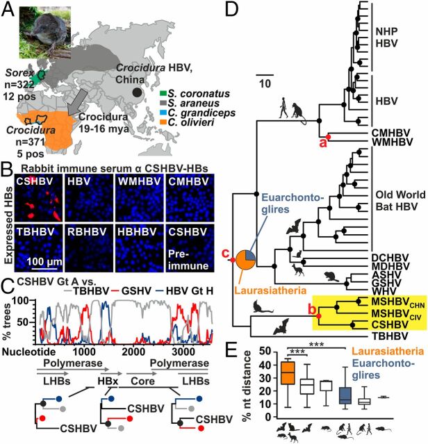
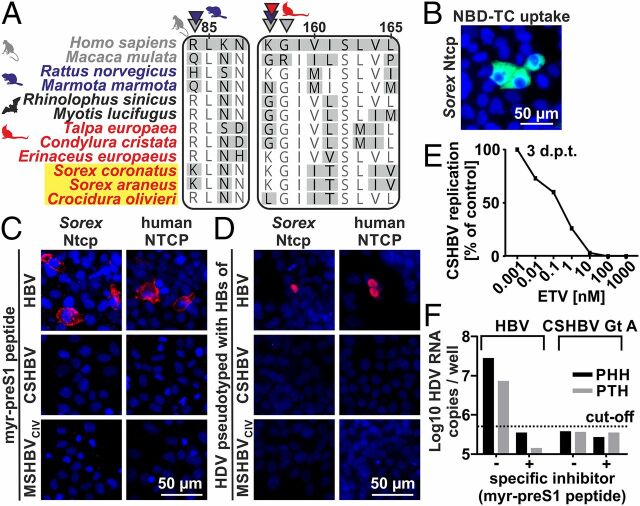
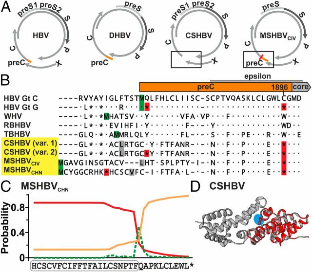
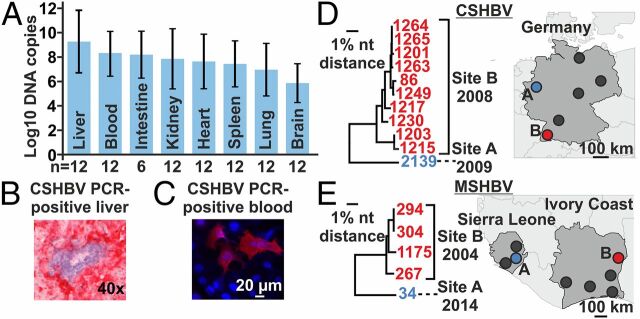
Shrews, insectivorous small mammals, pertain to an ancient mammalian order. We screened 693 European and African shrews for hepatitis B virus (HBV) homologs to elucidate the enigmatic genealogy of HBV. Shrews host HBVs at low prevalence (2.5%) across a broad geographic and host range. The phylogenetically divergent shrew HBVs comprise separate species termed crowned shrew HBV (CSHBV) and musk shrew HBV (MSHBV), each containing distinct genotypes. Recombination events across host orders, evolutionary reconstructions, and antigenic divergence of shrew HBVs corroborated ancient origins of mammalian HBVs dating back about 80 million years. Resurrected CSHBV replicated in human hepatoma cells, but human- and tupaia-derived primary hepatocytes were resistant to hepatitis D viruses pseudotyped with CSHBV surface proteins. Functional characterization of the shrew sodium taurocholate cotransporting polypeptide (Ntcp), CSHBV/MSHBV surface peptide binding patterns, and infection experiments revealed lack of Ntcp-mediated entry of shrew HBV. Contrastingly, HBV entry was enabled by the shrew Ntcp. Shrew HBVs universally showed mutations in their genomic preCore domains impeding hepatitis B e antigen (HBeAg) production and resembling those observed in HBeAg-negative human HBV. Deep sequencing and in situ hybridization suggest that HBeAg-negative shrew HBVs cause intense hepatotropic monoinfections and low within-host genomic heterogeneity. Geographical clustering and low MSHBV/CSHBV-specific seroprevalence suggest focal transmission and high virulence of shrew HBVs. HBeAg negativity is thus an ancient HBV infection pattern, whereas Ntcp usage for entry is not evolutionarily conserved. Shrew infection models relying on CSHBV/MSHBV revertants and human HBV will allow comparative assessments of HBeAg-mediated HBV pathogenesis, entry, and species barriers.

Keywords: E antigen; hepatitis B virus; shrew; viral evolution; zoonosis.


Conflict of interest statement

The authors declare no conflict of interest.

Figures

Fig. 1.
Fig. 1.
Evolutionary and antigenic characteristics of shrew HBVs. (A) HBV-positive shrew species, sample. Gray arrows, Soricidae dispersion routes (16). Sorex picture, Ulrike Rosenfeld. (B) Antigenic divergence of CSHBV. (C) Bootscan analysis with maximum likelihood (ML) phylogenies of highlighted genomic regions. Filled circles in A, B, and D, bootstrap support >90% (black) and >80% (gray). (D) Bayesian ancestral state reconstruction and hypothesis testing (42). Pie charts, posterior probabilities for ancestral traits. Filled circles at nodes, posterior probability >0.95. Red nodes, priors used for hypothesis testing (Scale bars, genetic distance). (E) Pairwise HBV nucleotide sequence distance comparisons per host order. Boxplots, interquartile range and median; whiskers, minimum to maximum. Gt, genotype; AA, amino acid; WMHBV, woolly monkey HBV; RBHBV, roundleaf bat HBV; HBHBV, horseshoe bat HBV; LHBs, large HBV surface protein; HBx, HBV X protein; NHP, nonhuman primates; DCHBV, domestic cat HBV; MDHBV, Maxwell’s duiker HBV; ASHV, arctic squirrel hepatitis virus; GSHV, ground squirrel hepatitis virus.
Fig. 2.
Fig. 2.
NTCP usage and zoonotic potential of shrew HBVs. (A) NTCP/Ntcp-preS1 binding sites. Arrows, sites under positive selection for primates (gray), rodents (blue) (27), and Eulipotyphla (red). Yellow background, shrew HBV hosts. (B) Taurocholate (NBD-TC) uptake (green) by Sorex Ntcp. Blue, nuclei. (C) Myristoylated preS1 peptide binding (red) to human or Sorex NTCP/Ntcp. (D) Infection of HDV pseudotypes via human or Sorex NTCP/Ntcp. Red, newly produced Delta antigen. Blue, nuclei. (E) Inhibition of CSHBV Gt A replication. ETV, Entecavir. (F) Infection of primary human/tupaia hepatocytes by HDV pseudotypes. d.p.t, days posttransfection; GE, genome equivalents; Gt, genotype.
Fig. 3.
Fig. 3.
Shrew HBV HBeAg. (A) Genome structures. (B) Translated preC and N-terminal core domains. Red, stop codons; green, methionine; gray, alternative start codons. Var. 1/2, HTS minority variants (25–30% occurrence). (C) Signal peptide prediction (red line) of MSHBVCHN. Green, cleavage site; orange line, no signal peptide; boxed, predicted signal sequence. (D) CSHBV HBeAg monomer (cyan, reverted G1896A) modeled on the HBV HBeAg dimer (43). Gt, genotype; RBHBV, roundleaf bat HBV.
Fig. 4.
Fig. 4.
Shrew HBV infection patterns. (A) Mean CSHBV concentrations in solid organs (grams) and blood (milliliters), SDs. (B) In situ hybridization of CSHBV. (C) Shrew serum reacting with CSHBV proteins. Red, antibody binding; blue, nuclei. (D and E) Major sampling sites in Germany, Sierra Leone, and Ivory Coast; neighbor-joining phylogenies. Trees encompass 773 (CSHBV) and 1,432 (MSHBV) nucleotides. One CSHBV strain for which only a smaller fragment was characterized is not shown.

Similar articles

Cited by

References

    1. Rasche A., Sander A. L., Corman V. M., Drexler J. F., Evolutionary biology of human hepatitis viruses. J. Hepatol. 70, 501–520 (2019). - PMC - PubMed
    1. Lauber C., et al. ., Deciphering the origin and evolution of hepatitis B viruses by means of a family of non-enveloped fish viruses. Cell Host Microbe 22, 387–399.e6 (2017). - PMC - PubMed
    1. Suh A., et al. ., Early mesozoic coexistence of amniotes and hepadnaviridae. PLoS Genet. 10, e1004559 (2014). - PMC - PubMed
    1. Foley N. M., Springer M. S., Teeling E. C., Mammal madness: Is the mammal tree of life not yet resolved? Philos. Trans. R. Soc. Lond. B Biol. Sci. 371, 20150140 (2016). - PMC - PubMed
    1. Gogarten J. F., et al. ., A novel orthohepadnavirus identified in a dead Maxwell’s Duiker (Philantomba maxwellii) in Taï National Park, Côte d’Ivoire. Viruses 11, E279 (2019). - PMC - PubMed

Publication types

Associated data

LinkOut - more resources