. 2019 Aug 20;116(34):16872-16881.
doi: 10.1073/pnas.1902843116. Epub 2019 Aug 1.

p120-catenin regulates WNT signaling and EMT in the mouse embryo

Affiliations

p120-catenin regulates WNT signaling and EMT in the mouse embryo

Rocío Hernández-Martínez et al. Proc Natl Acad Sci U S A. .

Abstract

Epithelial-to-mesenchymal transitions (EMTs) require a complete reorganization of cadherin-based cell-cell junctions. p120-catenin binds to the cytoplasmic juxtamembrane domain of classical cadherins and regulates their stability, suggesting that p120-catenin may play an important role in EMTs. Here, we describe the role of p120-catenin in mouse gastrulation, an EMT that can be imaged at cellular resolution and is accessible to genetic manipulation. Mouse embryos that lack all p120-catenin, or that lack p120-catenin in the embryo proper, survive to midgestation. However, mutants have specific defects in gastrulation, including a high rate of p53-dependent cell death, a bifurcation of the posterior axis, and defects in the migration of mesoderm; all are associated with abnormalities in the primitive streak, the site of the EMT. In embryonic day 7.5 (E7.5) mutants, the domain of expression of the streak marker Brachyury (T) expands more than 3-fold, from a narrow strip of posterior cells to encompass more than one-quarter of the embryo. After E7.5, the enlarged T+ domain splits in 2, separated by a mass of mesoderm cells. Brachyury is a direct target of canonical WNT signaling, and the domain of WNT response in p120-catenin mutant embryos, like the T domain, is first expanded, and then split, and high levels of nuclear β-catenin levels are present in the cells of the posterior embryo that are exposed to high levels of WNT ligand. The data suggest that p120-catenin stabilizes the membrane association of β-catenin, thereby preventing accumulation of nuclear β-catenin and excessive activation of the WNT pathway during EMT.

Keywords: WNT signaling; cell migration; epithelial–mesenchymal transition; gastrulation; p53-dependent cell death.


Conflict of interest statement

The authors declare no conflict of interest.

Figures

Fig. 1.
Fig. 1.
The posterior body axis is duplicated in p120-catenin mutant embryos. (AD) Expression of T in E8.5 wild-type and p120-catenin mutant embryos, assayed by in situ hybridization, dorsal views. (A) Wild-type embryos express T in the primitive streak and the midline. (B) Approximately 80% of p120-catenin–null mutants have a posterior bifurcation of the Brachyury (T) expression domain in the primitive streak (arrows point to the 2 T-domains). Arrowheads point to 2 allantoides. (C) Embryos homozygous for a truncating point mutation of p120-catenin (LX169) recapitulate the null phenotype. The arrowheads point to 2 allantoides. (D) Conditional deletion of p120-catenin in the epiblast. Most p120-ΔEpi mutants develop 2 allantoides (arrowheads). (EG) In situ hybridization for T in E7.5 wild-type and p120-catenin mutant embryos, posterior views. (E) T is expressed in the posterior of wild-type embryos. (F and G) T is expressed in 2 separated domains in the null (F) and mutant embryos with LX169 point mutation (G). The arrowheads point to 2 allantoides. (All scale bars, 50 μm.)
Fig. 2.
Fig. 2.
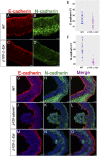
p120-catenin is required for normal Cadherin levels, but not for the E-cadherin to N-cadherin switch. (AD) High-magnification images of immunostained E7.5 transverse wild-type and mutant embryo sections stained for E-cadherin (red) and N-cadherin (green). (A and B) Wild type. (C and D) p120-ΔEpi mutants. (A) E-cadherin is concentrated in both apical and lateral membranes of the wild-type epiblast; (C) E-cadherin levels are reduced in the mutant, especially in lateral epiblast membranes. N-cadherin is expressed in the mesoderm layer of both (B) wild-type and (D) p120 mutant embryos, but the staining appears more diffuse and punctate in the mutant. (Scale bars, 16 μm.) (E) The E-cadherin fluorescence intensity (FI) of wild-type and mutant epiblast cells. The mean E-cadherin FI for the wild-type epiblast cells was 25.06 ± 1.57, whereas in the mutant FI = 16.50 ± 1.40; P < 0.0002 (n = 31 cells each for wild type and mutant). (F) N-cadherin FI for wild-type mesoderm cells was 41.67 ± 2.06; mesoderm mutant cells have a mean FI of 23.52 ± 0.71, P < 0.0001 (n = 23 cells each for wild type and mutant). Points are values for individual cells; bars represent the mean and SD. (GO) Higher-magnification views of cadherin expression in the streak region of E7.5 embryos. (GI) Wild-type embryo. (JL) A cluster of cells protruding from the mutant streak into the amniotic cavity expresses N-cadherin, and not E-cadherin. (MO) In other mutants, cells contiguous with the epiblast layer express N-cadherin. (Scale bars, 16 μm.)
Fig. 3.
Fig. 3.
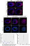
p120-catenin limits the size of the primitive streak and inhibits apoptosis during the EMT. (A and B) Immunostaining for T in E7.0 transverse section; posterior to the right. (A) Wild-type embryos show T in the posterior. (B) In the mutant epiblast, the single domain of T is expanded. (CG) Cryosections of E7.5 and E7.75 embryos stained for T. (AD) The arrows mark the edges of T-expression domain in the epiblast layer. (C and E) Wild-type embryos express T in the streak region of the posterior epiblast and in the nascent mesoderm derived from the streak. (D) In 80% of p120-catenin–null embryos (n = 12/15) and p120-ΔEpi (n = 8/10) mutant embryos, the domain of T expression in the posterior epiblast is expanded and split into 2 domains by T-negative cells. (F) In the remaining null and p120-ΔEpi mutants (n = 3/15 and n = 2/10), a cluster of T+ cells protrudes into the central amniotic cavity. The arrow points to pyknotic nuclei. The arrowhead points to nonspecific binding of T antibody to the visceral endoderm; compare with D using a different T antibody. (G) T expression in p120−/− p53−/− mutant embryos. (Scale bar, 50 μm.) (HJ) Sections of E7.5 embryos stained for Cleaved Caspase-3 (green). (H) Wild-type embryos do not have cell death. (J) p120−/− p53−/− double mutants have fewer apoptotic bodies than (I) p120 single mutants; speckles of Cleaved Caspase-3 were observed in p120 p53 double-mutant embryos (J). (All scale bars, 50 μm.) (K) Fraction of T-positive cells in the epiblast layer of wild-type and p120-catenin mutant embryos. Each dot represents the value from 1 section from 1 embryo; black bars represent the mean and SD. EPI, epiblast deleted; WT, wild type. In E7.5 wild-type embryos, 7.0% ± 0.52 of the cells in a single optical section were T+, whereas 11.4% ± 0.90 of the cells were T+ in E7.5 p120-ctn−/−, stained in the same experiment. In an independent experiment, 5.7 ± 0.67% of epiblast cells were T+ in E7.5 wild-type, whereas 8.9 ± 0.85% of cells were T+ in E7.5 p120-ΔEpi. At E7.0, 7.7% ± 0.63 of cells were T+ in wild type, whereas 10.9% ± 0.35 of cells were T+ cells in p120-ΔEpi embryos. In E7.5 p120 p53 double-mutant embryos 24.0± 4.20 were T+ cells, whereas 7.1 ± 0.89 were T+ in wild-type sections stained in parallel. The brackets indicate experiments stained in the same batch. (L) Quantitation of Cleaved Caspase-3 apoptotic bodies per section. Data are the mean ± SD (black bars). The mean number of Caspase-3+ apoptotic bodies detected per wild-type embryo was 0.66 ± 0.33; the mean was 46 ± 14 in p120−/− mutants; this number was reduced to 1.70 ± 1.2 in p120−/− p53−/− double mutants (n = 3 for each genotype; P < 0.0053).
Fig. 4.
Fig. 4.
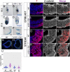
p120-catenin restricts the domain and level of WNT response in the posterior epiblast. (AH) WNT reporter gene expression. (A and B) β-Galactosidase staining for the Batgal WNT reporter in E6.5 (A) wild-type and (B) mutant embryos. Posterior to the right. (C and D) Topgal expression in E7.5 (C) wild-type and (D) p120-catenin–null embryos. Posterior view. (E and F) Transverse sections of E7.5 embryos stained for Batgal, (E) wild-type, and (F) p120-catenin mutants; posterior to the right. (G and H) Immunostaining for TCF-Lef:H2B-GFP-reporter expression in transverse sections of E7.5 embryos. The vertical white lines represent the extent of the GFP+ domain. (Scale bars, 50 μm.) (IU) Nuclear β-catenin. (IT) High-magnification views of immunostained E7.5 embryo sections, showing localization of β-catenin (white) and T (red). Upper panels correspond to the lateral epiblast of (IK) wild type and (LN) mutant; Lower panels show the streak of (OQ) wild type and (RT) mutant. Note colocalization β-catenin and T in nuclei of cells of the mutant streak. (Scale bar, 8 μm.) (U) Quantitation of fluorescence intensity (FI) of β-catenin in the nuclei of streak and epiblast cells (EPI). In wild-type embryos, the β-catenin FI in the nuclei at the streak was 15.65 ± 5.57; in contrast, in the mutant streak, the nuclear β-catenin FI was 35.86 ± 14.94 (P < 0.0001). The mean β-catenin FI in wild-type epiblast nuclei was 15.09 ± 7.49, whereas in the mutant epiblast was 14.45 ± 7.30 (P < 0.351). The black bars in the graphic represent the mean + SD.
Fig. 5.
Fig. 5.
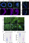
p120-catenin promotes persistent directional mesoderm migration. (AC) Immunostaining of E7.5 transverse embryo sections for N-cadherin (green) in (A) wild type, (B) p120-catenin, and (C) p53−/− p120−/− double mutants. (DF) Transverse sections of E7.5 embryos stained for SNAIL. (E) p120−/− and (F) p53−/− p120−/− double-mutant embryos have more SNAIL-expressing cells (magenta) in the epiblast layer than seen in (D) wild type. (D and E) The arrowheads point to nonspecific binding of SNAIL antibody to the visceral endoderm. (G and H) Immunofluorescent staining of transverse sections of E7.5 wild-type and p120-catenin mutant embryos for KDR (green). (Scale bars, 50 μm.) N-cadherin expression in mesoderm explants of (IK) wild-type and (LN) p120-ΔEpi mutant embryos. (Scale bars: I and L, 80 μm; J and M, 24 μm; K and N, 8 μm.) (O) The average velocity of migration of wild-type and mutant mesoderm cell explants over a 7-h period of culture. Points are values for individual cells; bars represent the mean and SD. The mean velocity for the mutant explants was 0.48 ± 0.02 μm/min, 70% faster than wild type (0.28 ± 0.01 μm/min; P < 0.0001) (n = 54 cells each for wild type and mutant). (P) Comparison of the cell migration directionality (persistence) in wild-type and mutant mesoderm cell explants over a 7-h period of culture. Data are shown as the mean persistence and SD for wild-type (0.78 ± 0.02) and mutant (0.36 ± 0.03) mesoderm cells (P < 0.0001). A value of 1.0 is a constant direction.
Fig. 6.
Fig. 6.
Posterior axis splitting and tissue disorganization in p120 p53 double-mutant embryos. (AC) Whole-mount in situ hybridization of E8.5 embryos for T expression. (A) T expression in the wild-type embryo. (B) The 2-T main domains seem connected by T-expressing cells, and there are 2 allantoides in p120−/−p53−/− and (C) p120-ΔEpi p53-ΔEpi mutant embryos. The arrowheads point to 2 allantoides. (Scale bar, 50 μm.) (DO) Primitive streak of E8.5 wild-type and p120 p53 double-mutant embryos. (D and J) In wild-type, T (magenta) is expressed in the streak and in the nascent mesoderm. (E) N-cadherin (green) marks mesoderm cells and cadherin switching has started in the apical epiblast. (F) E-cadherin (red) is expressed throughout the epiblast. In the majority of p120 p53 double-mutant embryos examined at this stage (3/5), (G) T is expressed in 2 domains, (H) separated by a mass of cells expressing punctate N-cadherin and not covered by (I) E-cadherin–expressing epiblast. In the other E8.5 double mutants (MO), the tissue in the posterior of the double mutants is disorganized, with regions expressing SNAIL (green) and CD31 (red) (N and O). (Scale bar, 50 μm.)

Similar articles

Cited by

References

    1. Ishiyama N., et al. , Dynamic and static interactions between p120 catenin and E-cadherin regulate the stability of cell-cell adhesion. Cell 141, 117–128 (2010). - PubMed
    1. Nanes B. A., et al. , p120-catenin binding masks an endocytic signal conserved in classical cadherins. J. Cell Biol. 199, 365–380 (2012). - PMC - PubMed
    1. Myster S. H., Cavallo R., Anderson C. T., Fox D. T., Peifer M., Drosophila p120catenin plays a supporting role in cell adhesion but is not an essential adherens junction component. J. Cell Biol. 160, 433–449 (2003). - PMC - PubMed
    1. Pettitt J., Cox E. A., Broadbent I. D., Flett A., Hardin J., The Caenorhabditis elegans p120 catenin homologue, JAC-1, modulates cadherin-catenin function during epidermal morphogenesis. J. Cell Biol. 162, 15–22 (2003). - PMC - PubMed
    1. Bulgakova N. A., Brown N. H., Drosophila p120-catenin is crucial for endocytosis of the dynamic E-cadherin-Bazooka complex. J. Cell Sci. 129, 477–482 (2016). - PMC - PubMed

Publication types