. 2019 Sep 3;93(10):e954-e963.
doi: 10.1212/WNL.0000000000008061. Epub 2019 Aug 1.

Autoimmune gait disturbance accompanying adaptor protein-3B2-IgG

Affiliations

Autoimmune gait disturbance accompanying adaptor protein-3B2-IgG

Josephe A Honorat et al. Neurology. .

Erratum in

Abstract

Objective: To describe phenotypes, treatment response, and outcomes of autoimmunity targeting a synaptic vesicle coat protein, the neuronal (B2) form of adaptor protein-3 (AP3).

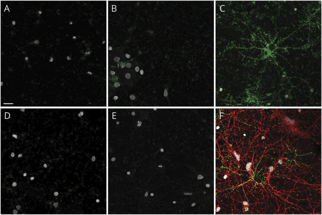
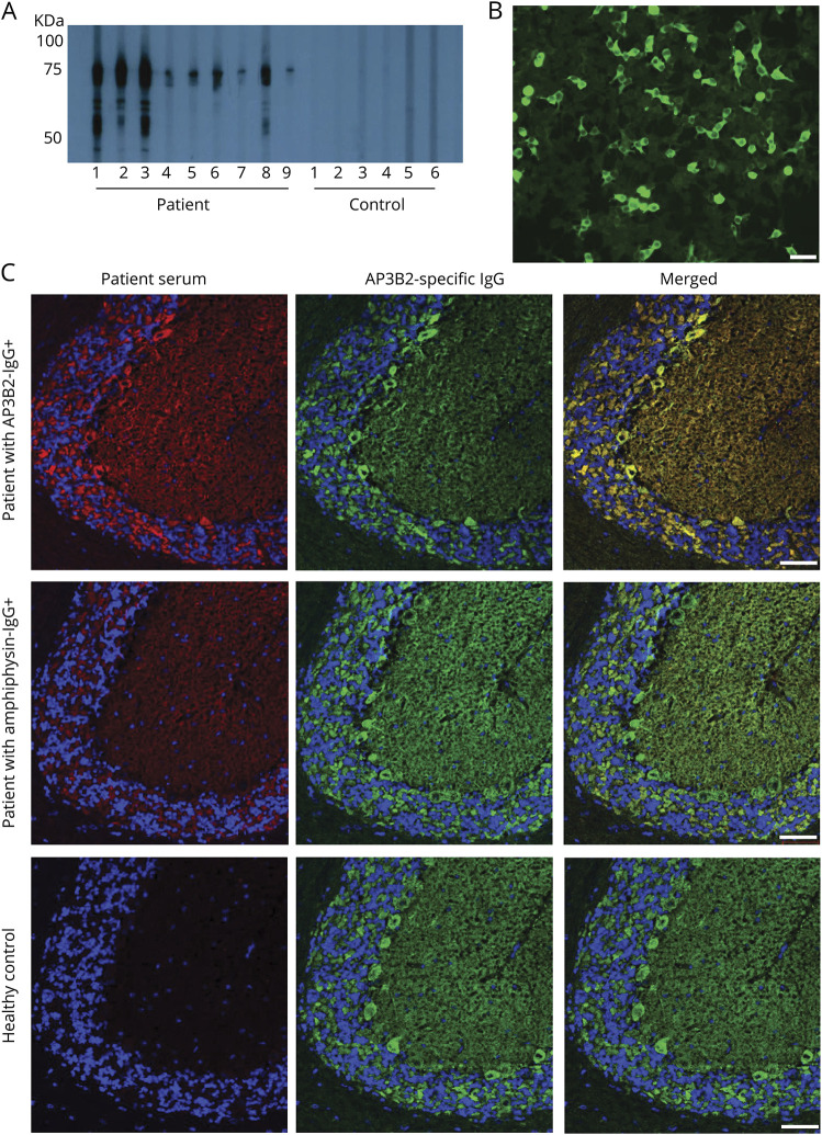
Methods: Archived serum and CSF specimens (from 616,025 screened) harboring unclassified synaptic antibodies mimicking amphiphysin-immunoglobulin G (IgG) on tissue-based indirect immunofluorescence assay (IFA) were re-evaluated for novel IgG staining patterns. Autoantigens were identified by western blot and mass spectrometry. Recombinant western blot and cell-binding assay (CBA) were used to confirm antigen specificity. Clinical data were obtained retrospectively.

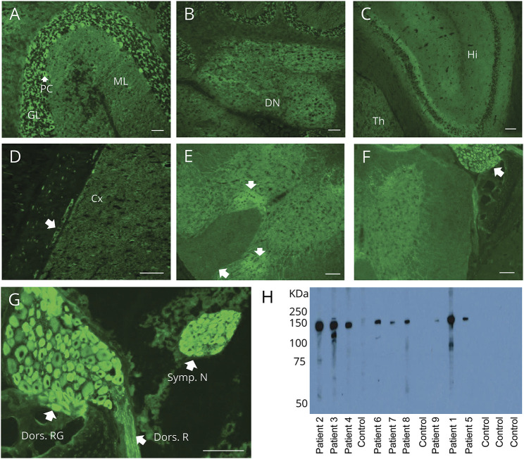
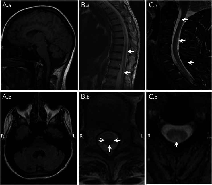
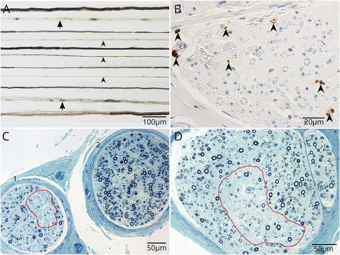
Results: Serum (10) and CSF (6) specimens of 10 patients produced identical IFA staining patterns throughout mouse nervous system tissues, most prominently in cerebellum (Purkinje neuronal perikarya, granular layer synapses, and dentate regions), spinal cord gray matter, dorsal root ganglia, and sympathetic ganglia. The antigen revealed by mass spectrometry analysis and confirmed by recombinant assays (western blot and CBA) was AP3B2 in all. Of 10 seropositive patients, 6 were women; median symptom onset age was 42 years (range 24-58). Clinical information was available for 9 patients, all with subacute onset and rapidly progressive gait ataxia. Neurologic manifestations were myeloneuropathy (3), peripheral sensory neuropathy (2), cerebellar ataxia (2), and spinocerebellar ataxia (2). Five patients received immunotherapy; none improved, but they did not worsen over the follow-up period (median 36 months; range 3-94). Two patients (both with cancer) died. One of 50 control sera was positive by western blot only (but not by IFA or CBA).

Conclusion: AP3B2 (previously named β-neuronal adaptin-like protein) autoimmunity appears rare, is accompanied by ataxia (sensory or cerebellar), and is potentially treatable.


Figures

Figure 1
Figure 1. Distribution of adaptor protein 3, subunit B2 immunoreactivity in mouse tissues revealed by patient immunoglobulin G (IgG) binding
(A–G) Patient serum IgG yields synaptic pattern of immunofluorescence in (A) cerebellar granular layer (GL) more than molecular layer (ML), plus perikarya of Purkinje cells (PC); (B) cerebellar dentate nucleus (DN); (C) hippocampus (Hi), thalamus (Th); (D) cerebral cortex (Cx) and ganglionic neuronal perikarya in juxtaposed gastric smooth muscle (arrow); and (E) spinal cord gray matter (particularly dorsal regions [arrows]). (F, G) Immunoreactivity is most prominent in perikarya of dorsal root (F, enlarged in G) and sympathetic ganglionic neurons and dorsal root nerve fibers (G). (H) In western blot, cytosolic lysate of mouse cerebellum contains a prominent antigenic protein of approximately 150 kDa, reactive with IgG in sera of patients 1–9, but not with healthy control sera IgG. Scale bar: A, B, D, 50 μm; C, E–G: 100 μm.
Figure 2
Figure 2. Live cell binding of control and patient immunoglobulin G (IgG) on hippocampal neurons
(A) Control CSF from normal pressure hydrocephalus patient and (B) healthy control serum do not bind to the surface of living hippocampal neurons. Neither CSF (D) nor serum (E) from patients 1–10 (representative images from patient 5) bind to the neuronal cultures. In contrast, NMDAR-IgG-positive patient CSF binds in a punctate pattern to the extracellular surface of hippocampal neurons (C, green). Cells were poststained for acetylated tubulin to identify axons (F, red). Nuclei stained with DAPI in all panels (white). Scale bar, 20 μm.
Figure 3
Figure 3. Western blot with recombinant protein and confocal microscopy confirm the target antigen is a cytosolic protein, adaptor protein 3, subunit B2 (AP3B2)
(A) By western blot, immunoglobulin G (IgG) from 9 patient sera, but none of 6 healthy control sera shown, bind to a recombinant AP3B2 C-terminal polypeptide fragment. (B) Positivity by AP3B2-specific cell-binding assay in serum of patient 7. (C) Commercial AP3B2 antibody colocalizes well with immunoreactivity produced on mouse cerebellum by patient AB3B2-IgG (top), but not with amphiphysin-IgG-positive patient (middle) or healthy control (bottom). Left column, patient IgG; middle column, rabbit anti‐AP3B2 IgG; right column, merged images. Scale bars, 50 μm.
Figure 4
Figure 4. Brain and spinal cord MRI
Patient 1: (A.a) Prominent cerebellar atrophy in sagittal T1-weighted brain MRI. (A.b) Enlarged IVth ventricle in axial T1-weighted brain MRI. Patient 3: (B.a) Longitudinal lesion extending from T9 to conus medullaris in sagittal T2-weighted thoracolumbar spine MRI. (B.b) Increased T2 signal in spinal cord gray matter and posterior columns (arrows) consistent with tractopathy (axial T2-weighted). Patient 5: (C.a, C.b) Increased T2 signal in spinal cord posterior columns (arrows) in sagittal (C.a) and axial (C.b) T2-weighted cervical spine MRI.
Figure 5
Figure 5. Whole sural nerve biopsy of patient 5 demonstrates multifocal axonal degeneration without evident inflammation
(A) Closely approximated teased nerve fibers stained with osmium tetroxide show increased empty nerve strands (arrowheads) and increased axonal degeneration (arrows). (B) Formalin-fixed paraffin section. CD68 immunoreactive cells are more numerous than normal; presumed macrophages, but lacking CD45 immunoreactivity. (C, D) Loss of axonal fibers is variable between fascicles and within fascicles (C, D, red outlined).

References

    1. McKeon A, Pittock SJ. Paraneoplastic encephalomyelopathies: pathology and mechanisms. Acta Neuropathol 2011;122:381–400. - PubMed
    1. Dalmau J, Graus F, Rosenblum MK, Posner JB. Anti-Hu-associated paraneoplastic encephalomyelitis/sensory neuronopathy: a clinical study of 71 patients. Medicine 1992;71:59–72. - PubMed
    1. Lucchinetti CF, Kimmel DW, Lennon VA. Paraneoplastic and oncologic profiles of patients seropositive for type 1 antineuronal nuclear autoantibodies. Neurology 1998;50:652–657. - PubMed
    1. Darnell RB, Furneaux HM, Posner JB. Antiserum from a patient with cerebellar degeneration identifies a novel protein in Purkinje cells, cortical neurons, and neuroectodermal tumors. J Neurosci 1991;11:1224–1230. - PMC - PubMed
    1. Honorat JA, Lopez-Chiriboga AS, Kryzer TJ, et al. Autoimmune septin-5 cerebellar ataxia. Neurol Neuroimmunol Neuroinflamm 2018;5:e474. - PMC - PubMed

Publication types

MeSH terms