. 2019 Aug 2;10(8):579.
doi: 10.1038/s41419-019-1818-4.

Site-specific phosphorylation of Fbxw7 by Cdk5/p25 and its resulting decreased stability are linked to glutamate-induced excitotoxicity

Affiliations

Site-specific phosphorylation of Fbxw7 by Cdk5/p25 and its resulting decreased stability are linked to glutamate-induced excitotoxicity

Yeon Uk Ko et al. Cell Death Dis. .

Abstract

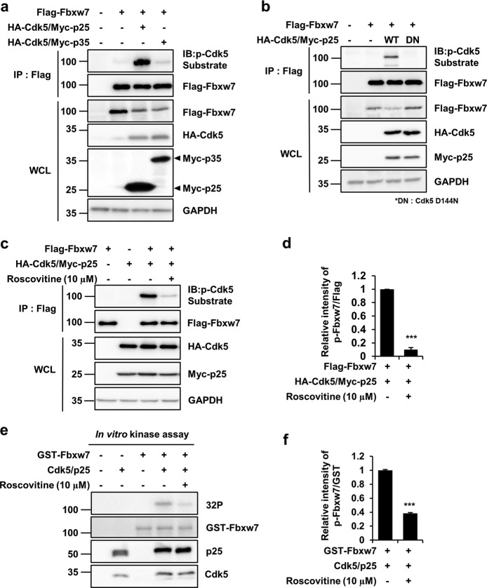
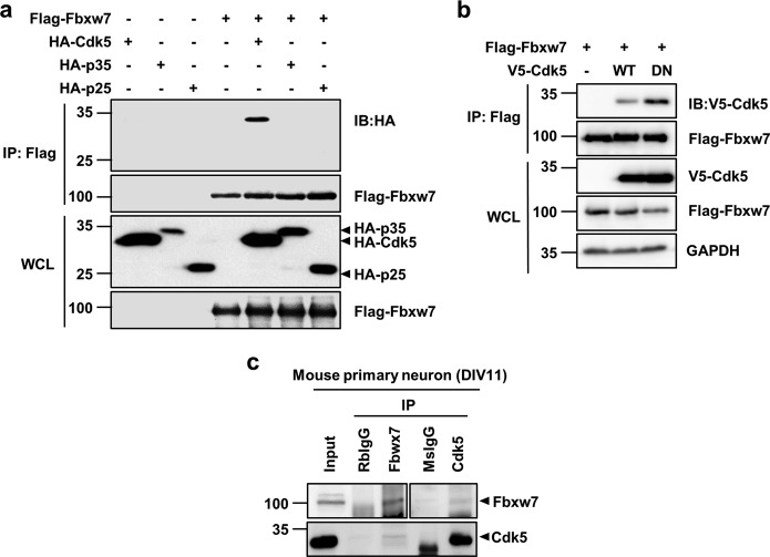
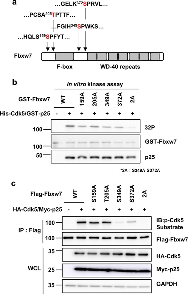
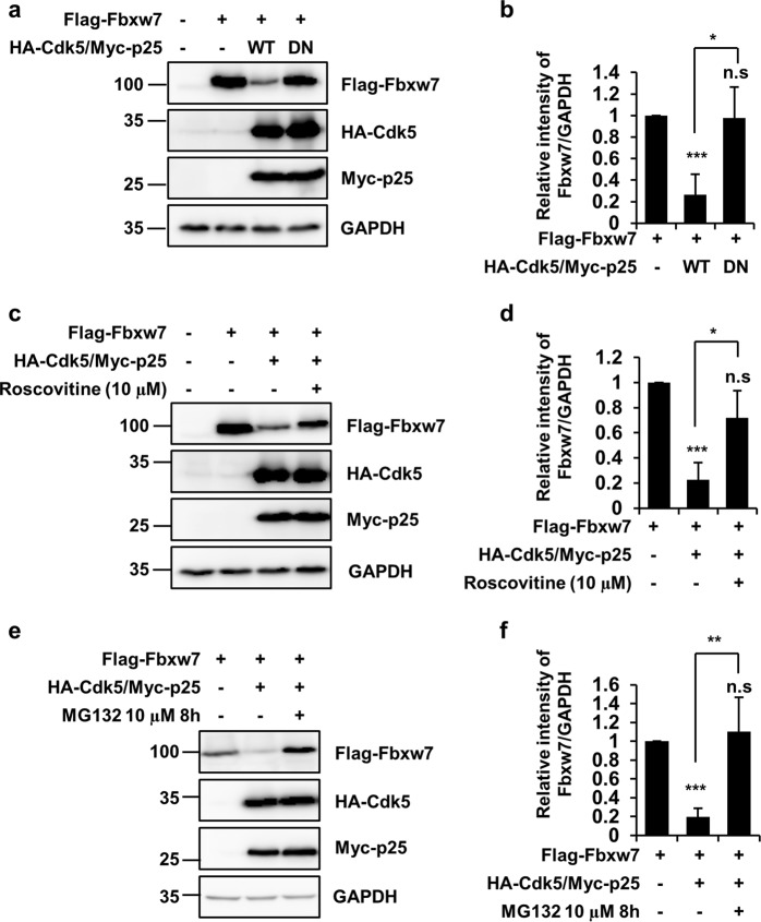
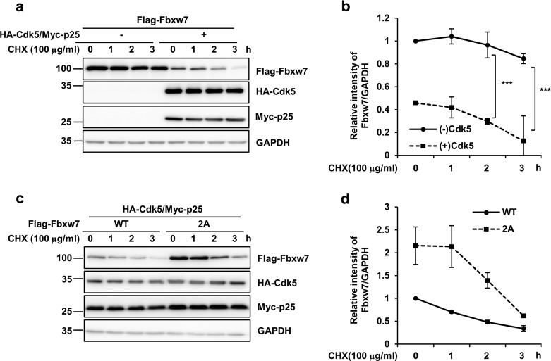
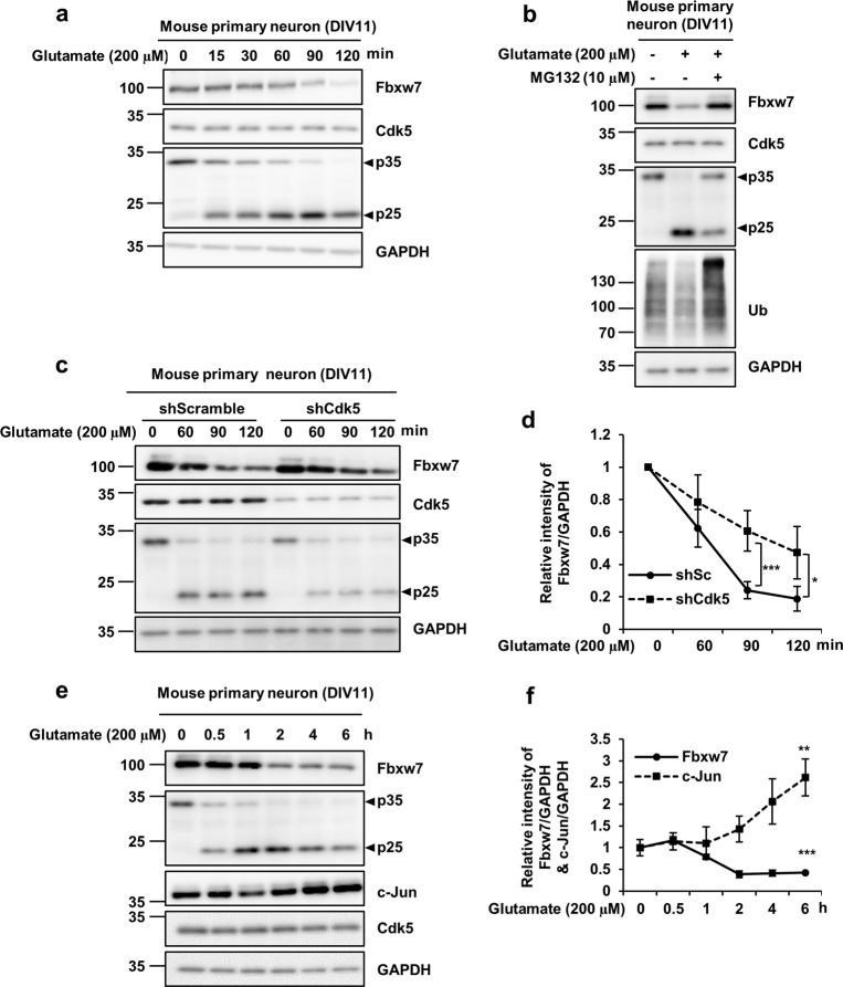
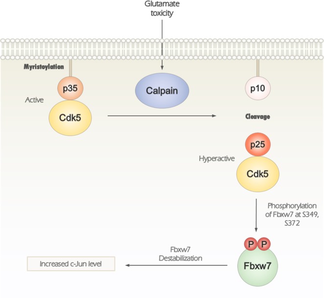
Cyclin-dependent kinase 5 (Cdk5) is a serine/threonine protein kinase that regulates brain development and neurodegeneration. Cdk5 is activated by p25 that is generated from calpain-dependent cleavage of p35. The generation of p25 is responsible for the aberrant hyper-activation of Cdk5, which causes neurodegeneration. Using in vitro assays, we discovered that F-box/WD repeat-containing protein 7 (Fbxw7) is a new substrate of Cdk5. Additionally, Cdk5-dependent phosphorylation of Fbxw7 was detected in the presence of p25, and two amino acid residues (S349 and S372) were determined to be major phosphorylation sites. This phosphorylation was eventually linked to decreased stability of Fbxw7. Using a culture model of cortical neurons challenged with glutamate, we confirmed that decreased stability of Fbxw7 was indeed Cdk5-dependent. Furthermore, diminished levels of Fbxw7 led to increased levels of transcription factor AP-1 (c-Jun), a known substrate of Fbxw7. Given that previous reports demonstrate that c-Jun plays a role in accelerating neuronal apoptosis in these pathological models, our data support the concepts of a molecular cascade in which Cdk5-mediated phosphorylation of Fbxw7 negatively regulates Fbxw7 expression, thereby contributing to neuronal cell death following glutamate-mediated excitotoxicity.


Conflict of interest statement

The authors declare that they have no conflict of interest.

Figures

Fig. 1
Fig. 1. Cdk5/p25 phosphorylates Fbxw7.
a HEK293 cells were transfected with Flag-Fbxw7 in combination with either HA-Cdk5/Myc-p25, HA-Cdk5/Myc-p35, or neither for the control. Cell lysates were incubated with Flag-conjugated beads for immunoprecipitation (IP). Immunoprecipitates and whole cell lysates (WCL) were resolved by SDS-PAGE and followed by immunoblot analysis (IB). Phospho-Fbxw7 signal was visualized with an anti-phospho-Cdk5-substrate antibody. GAPDH was used as a loading control. b HEK293 cells were transfected with Flag-Fbxw7 in combination with either HA-Cdk5 WT/Myc-p25 or HA-Cdk5 dominant-negative D144N (DN)/Myc-p25. WCL were used to IP using Flag-conjugated beads and IB analysis was done using the indicated antibodies. c HEK293 cells were exposed to 10 μM roscovitine for 48 h. Immunoprecipitates were purified with an anti-Flag antibody and IB was conducted with the indicated antibodies. d After normalization to Flag-Fbxw7, the relative fold intensity of the phospho-Fbxw7 signal by Cdk5/p25 were measured in samples treated with or without roscovitine (value = 1). Bar represents the mean ± SD of three independent experiments. ***p < 0.001. e Purified GST-Fbxw7 protein (1 µg) was incubated with or without 0.1 µg recombinant His-Cdk5/GST-p25 protein in the presence of [γ-32P] ATP. Reaction mixtures were resolved by SDS-PAGE and subjected to autoradiography. f After normalization to GST-Fbxw7, the relative fold intensity of the phospho-Fbxw7 signal by Cdk5/p25 was measured in samples treated with or without roscovitine (value = 1). The bar represents the mean ± SD of three independent experiments. ***p < 0.001
Fig. 2
Fig. 2. Cdk5 interacts with Fbxw7.
a Following transfection of HEK293 cells with the indicated combination of vectors, cell lysates were subjected to IP using Flag-conjugated beads. Immunoprecipitates and WCL were resolved by SDS-PAGE and analyzed by IB analysis. b HEK293 cells were transfected with Flag-Fbxw7 alone or in combination with either V5-Cdk5 WT/Myc-p25 or V5-Cdk5 DN/Myc-p25. IP followed by IB was performed as described above. c Primary cultures of cortical neurons were maintained until days in vitro(DIV) 11. IP was performed with anti-Fbxw7 or anti-Cdk5 antibodies. Rabbit anti-IgG and mouse anti-IgG antibodies were used as IP controls. Fbxw7 was detected by IB after IP pulldown with anti-Cdk5 antibody and vice versa
Fig. 3
Fig. 3. Both Ser349 and Ser372 are the major sites of Fbxw7 that are phosphorylated by Cdk5/p25.
a Scheme for putative Cdk5-phosphorylaton sites of Fbxw7 (S/TPXK/R). b In vitro kinase assay to detect the direct phosphorylation sites of Fbxw7 by Cdk5/p25. Purified GST-Fbxw7 wild-type (WT) protein (1 µg) or one of the GST-Fbxw7 site-specific mutant proteins were incubated with 0.1 µg recombinant His-Cdk5/GST-p25 protein in the presence of [γ-32P] ATP. Reaction mixtures were resolved by SDS-PAGE and subjected to autoradiography. c HEK293 cells were transfected with Flag-Fbxw7 WT or one of the Flag-Fbxw7 site-specific mutant proteins in the presence or absence of HA-Cdk5/Myc-p25. For comparison with the p-Fbxw7 signal, HEK293 cells were transfected with Flag-Fbxw7 2A mutant plus HA-Cdk5/Myc-p25. Cell lysates were incubated with Flag-conjugated beads for IP. Immunoprecipitates and WCL were resolved by SDS-PAGE analyzed by IB analysis. Phospho-Fbxw7 signal was visualized with an anti-phospho-Cdk5-substrate antibody
Fig. 4
Fig. 4. Cdk5 negatively regulates Fbxw7 protein expression level.
a HEK293 cells were transfected with Flag-Fbxw7 alone or in combination with either HA-Cdk5 WT/Myc-p25 or HA-Cdk5 DN/Myc-p25. IB analysis was conducted using the indicated antibodies. b Relative intensity of Flag-Fbxw7 was calculated over the control (only transfected with Fbxw7; value = 1) after normalization against GAPDH. Bar represents the mean ± SD of three independent experiments. *p < 0.05; ***p < 0.001; n.s not significant. c HEK293 cells were exposed to 10 μM roscovitine for 48 h. IB analysis was conducted with the indicated antibodies. d Relative intensity of Flag-Fbxw7 intensity was calculated over the control (value = 1) after normalization against GAPDH. Bar represents the mean ± SD of three independent experiments. *p < 0.05; ***p < 0.001; n.s not significant. e HEK293 cells transfected with Flag-Fbxw7 alone or in combination with HA-Cdk5/Myc-p25 were treated with or without 10 μM MG132 for 8 h. Cell lysates were analyzed by IB using the indicated antibodies. f Relative intensity of Flag-Fbxw7 was calculated over the control (value = 1) after normalization against GAPDH. Bar represents the mean ± SD of three independent experiments. **p < 0.01; ***p < 0.001; n.s not significant
Fig. 5
Fig. 5. Cdk5-mediated phosphorylation causes the decreased stability of Fbxw7.
a HEK293 cell were transfected with Flag-Fbxw7 WT alone or in combination with Cdk5/Myc p25. Forty-eight hour after transfection, cells were incubated in the presence of 100 μg/ml cycloheximide (CHX) for the indicated time periods. Cell lysates from each condition were analyzed by IB using the indicated antibodies. b After normalization against GAPDH, relative levels of Flag-Fbxw7 at the designated time periods were evaluated against time point 0 and shown in a graph. Bar represents the mean ± SD of three independent experiments. ***p < 0.001. c HEK293 cells were transfected with Flag-Fbxw7 WT or Flag-Fbxw7 2A plus HA-Cdk5/Myc-p25. Forty-eight hour after transfection, cells were incubated with 100 μg/ml CHX. Cell lysates were analyzed by IB using the indicated antibodies. d After normalization against GAPDH, relative levels of Flag-Fbxw7 at the designated time periods were evaluated against time point 0 and shown in a graph. Bar represents the mean ± SD of three independent experiments
Fig. 6
Fig. 6. Inverse expression patterns of Fbxw7 and its substrate c-Jun detected in cortical neurons during glutamate excitotoxicity.
a Primary cultures of cortical neurons were exposed to 200 μM glutamate for the indicated time periods or b cultures were exposed for 2 h to 200 μM glutamate in the presence or absence of 10 μM MG132. a, b Cell lysates were harvested and analyzed by IB. c Primary cultures of cortical neurons were infected with lentiviral particles containing shRNA against either Cdk5 or control (Scramble) at DIV3 and subsequently maintained until DIV 11. Cultures were then exposed to 200 μM glutamate for the indicated time periods. Cell lysates were subjected to IB analysis. d After normalization against GAPDH, relative levels of Fbxw7 at the designated time periods were evaluated against time point 0 and are shown in the graph. Each point represents the mean ± SD from five independent experiments. *p < 0.05; ***p < 0.001. e Primary cultures of cortical neurons were exposed to 200 μM glutamate for up to 6 h. Cell lysates were harvested and analyzed by IB. f After normalization against GAPDH, relative fold intensity of Fbxw7 and c-Jun signal was quantified over the control at time 0 (value = 1). Each point represents the mean ± SD from three independent experiments. **p < 0.01; ***p < 0.001
Fig. 7
Fig. 7. A schematic model of the consequence of Cdk5-mediated phosphorylation of Fbxw7.
During glutamate-mediated excitotoxicity, activated calpain cleaves p35 into p25 leading to Cdk5 hyper-activation. Cdk5 in association with of activating cofactor p25 phosphorylates Fbxw7 at S349 and S372. Subsequently, Cdk5/p25-mediated phosphorylation induces destabilization of Fbxw7, resulting in a concomitant increase of its cellular substrate, c-Jun

References

    1. Morgan DO. Cyclin-dependent kinases: engines, clocks, and microprocessors. Annu. Rev. Cell Dev. 1997;13:261–291. doi: 10.1146/annurev.cellbio.13.1.261. - DOI - PubMed
    1. Satyanarayana A, Kaldis P. Mammalian cell-cycle regulation: several Cdks, numerous cyclins and diverse compensatory mechanisms. Oncogene. 2009;28:2925–2939. doi: 10.1038/onc.2009.170. - DOI - PubMed
    1. Dhavan R, Tsai LH. A decade of CDK5. Nat. Rev. Mol. Cell Biol. 2001;2:749–759. doi: 10.1038/35096019. - DOI - PubMed
    1. Kawauchi T. Cdk5 regulates multiple cellular events in neural development, function and disease. Dev. Growth Differ. 2014;56:335–348. doi: 10.1111/dgd.12138. - DOI - PubMed
    1. Malumbres M. Cyclin-dependent kinases. Genome Biol. 2014;15:122. doi: 10.1186/gb4184. - DOI - PMC - PubMed

Publication types

MeSH terms