. 2019 Aug 1;10(1):3446.
doi: 10.1038/s41467-019-11399-z.

Identification of a neurocircuit underlying regulation of feeding by stress-related emotional responses

Affiliations

Identification of a neurocircuit underlying regulation of feeding by stress-related emotional responses

Yuanzhong Xu et al. Nat Commun. .

Abstract

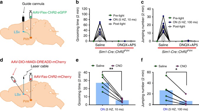
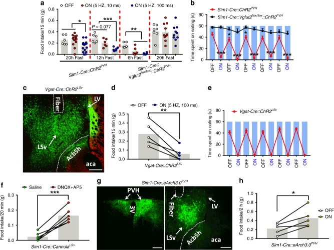
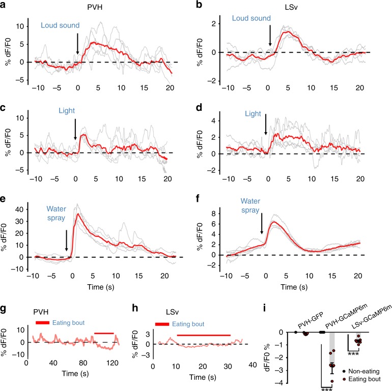
Feeding is known to be profoundly affected by stress-related emotional states and eating disorders are comorbid with psychiatric symptoms and altered emotional responses. The neural basis underlying feeding regulation by stress-related emotional changes is poorly understood. Here, we identify a novel projection from the paraventricular hypothalamus (PVH) to the ventral lateral septum (LSv) that shows a scalable regulation on feeding and behavioral changes related to emotion. Weak photostimulation of glutamatergic PVH→LSv terminals elicits stress-related self-grooming and strong photostimulation causes fear-related escape jumping associated with respective weak and strong inhibition on feeding. In contrast, inhibition of glutamatergic inputs to LSv increases feeding with signs of reduced anxiety. LSv-projecting neurons are concentrated in rostral PVH. LSv and LSv-projecting PVH neurons are activated by stressors in vivo, whereas feeding bouts were associated with reduced activity of these neurons. Thus, PVH→LSv neurotransmission underlies dynamic feeding by orchestrating emotional states, providing a novel neural circuit substrate underlying comorbidity between eating abnormalities and psychiatric disorders.


Conflict of interest statement

The authors declare no competing interests.

Figures

Fig. 1
Fig. 1
A local GABAergic network in the ventral part of lateral septum (LSv) receives direct glutamatergic input from PVH neurons. ac Sim1-Cre reporter mice (four males and two females) received injections of AAV-Flex-ChR2-eGFP vectors to one side of the PVH a, facilitating dual expression of the ROSA-lsl-tdTomato allele in Sim1-Cre neurons, and unilateral targeted expression of the AAV-Flex-ChR2-eGFP b. c GFP-expressing fibers from the PVH shown in the ventral part of lateral septum (LSv) were observed 4 weeks after AAV-delivery. dk Patch clamp recording of randomly selected neurons in LSv in brain slices from Sim1-Cre dj or Sim1-Cre::Vglut2flox/flox mice k with photostimulation of PVH→LSv fibers. df Voltage–clamp recordings for photostimulated (1 ms, blue ticks) excitatory postsynaptic currents (EPSCs) and their responses to glutamate receptor antagonists (CNQX+APV) d, GABA-A receptor antagonists (GABAzine) e, and 4-AP+TTX to block action potentials f. gj Voltage–clamp recordings with photostimulation (1 ms, blue ticks) to monitor inhibitory postsynaptic currents (oIPSCs) and their responses to GABAzine g, CNQX+APV h, 4-AP+TTX i and j. i and j showed the recording from the same set of neurons with complete i or partial blockage j by 4-AP/TTX. k Only three out of 25 recorded neurons showed 4-AP/TTX-resistant IPSC in Sim1-Cre::Vglut2flox/flox mice. The ratios at the top of traces indicate the number of neurons that exhibited responses to drugs out of all neurons showing postsynaptic currents. l A diagram derived from the recording data depicting a GABAergic network in the LSv receiving monosynaptic projections from the PVH comprising a major glutamatergic, and minor GABAergic components. Scale bar = 100 µm. PVH: paraventricular hypothalamus; LSv: ventral part of lateral septum; MS; medial septum; HDB and VDB: diagonal band (horizontal and vertical); LV: lateral ventricle; 3 V: the third ventricle; AcbSh: accumbens shell
Fig. 2
Fig. 2
Activation of PVH→LSv projections drives behaviors associated with negative emotional states. ac Sim1-Cre and Sim1-Cre::Vglut2flox/flox mice (males) with AAV-Flex-ChR2-eGFP delivery to the PVH and fiber optic implantation targeting LSv were used for behavioral assays a. Representative pictures showing AAV vector expression in the PVH b, arrows, and GFP-expressing fibers and optic fiber trace in LSv c, arrows. df These mice were used for a 2 min testing period. In Sim1-Cre mice, self-grooming behaviors were induced by short-pulse phototimulation (10 ms/5 Hz, 5 mW/mm2) (d, online Movie 1) and frantic jumping behaviors were induced by long pulse photostimulation (100 ms/5 Hz, 5 mW/mm2) (f, online Movie 2), neither of which were observed in Sim1-Cre::Vglut2flox/flox mice d and f. e Quantitation of self-grooming behaviors shows increased bout and transition (n = 10–11; comparison between % incorrect transitions, p < 0.001; comparison between % interrupted bouts, p = 0.012). g, h Mice were used for a shelter box test in which mice movements were tracked in an open arena with box (shaded areas in g) placed in the center. More time spent in the box represents an increase in fear. Photostimulation of PVH→LSv terminals increased time spent in the box, or in the same area the box occupied, compared with either photostimalation without the box or without photostimulation (h, n = 4, p = 0.0108 or p = 0.0113). i Representative snapshot pictures of attacking intruders by resident mice with no (OFF) or the long pulse photostimulation (ON) of PVH→LSv fibers (top pictures), and time spent in attack during the 30 s periods of pre-light and light-on (ON, also online Movie 3), and 2 mins post light (i, bottom; n = 11, p < 0001). *p < 0.05, ***p < 0.001, paired student’s t tests for e and I, One-way AVONA tests for h. Scale bar = 100 µm. PVH: paraventricular hypothalamus; LSv: ventral part of lateral septum; 3 V: the third ventricle; aca: anterior commissure area; AcbSh: accumbens shell
Fig. 3
Fig. 3
LSv neurons mediate the effect of activation of PVH→LSv. ac The effect of blocking LSv glutamate receptors on behavior. A paradigm depicting the experimental strategy using male Sim1-Cre mice a and the effect on self-grooming b and jumping c by infusion of saline or glutamate receptor antagonists DNQX+AP5 to LSv 5 mins prior to behavioral testing. df Vgat-Cre male mice with AAV-Fas-ChR2-eGFP (“Cre-off”) delivery to PVH, AAV-DIO-hM4Di-EREADD-mCherry delivery to LSv, and optic fiber implantation targeting LSv d were used for self-grooming (e n = 5, p = 0.0021) and jumping assays (f, n = 5, p = 0.0105) in response to saline and CNO treatment. PVH: paraventricular hypothalamus; LSv: ventral part of lateral septum; aca: anterior commissure area; 3 V: the third ventricle; AcbSh: accumbens shell. *p < 0.05, ** p < 0.01, paired student’s t tests e, f
Fig. 4
Fig. 4
Photostimulation of PVH→LSv terminals produces negative valence. ae Real time place preference were conducted in male mice with photostimulation of PVH→LSv fibers. A representative mouse locomotion trace was shown with short a and long b pulse photostimulation in Sim1-Cre mice. Time spent during laser on (ON) and off zones (OFF) in Sim1-Cre and Sim1-Cre::Vglut2flox/flox mice with AAV-Flex-ChR2-eGFP delivery, or Sim1-Cre mice with AAV-Flex-GFP delivery using the short pulse (c, Sim1-Cre::ChR2PVH mice n = 7, p = 0.0321) and the long pulse stimulation (d, Sim1-Cre::ChR2PVH mice n = 12, p < 0.0001). e Time spent in OFF zone shows dependence on the photostimulation duration with fixed 5 Hz and 5 mW/mm2 light (n = 5; 5 HZ,10 ms vs 5 HZ,50 ms, p = 0.0452; 5 HZ,50 ms vs 5 HZ,100 ms, p = 0.0356; 5 HZ,10 ms vs 5 HZ,50 ms, p < 0.0001). fh Mice described in ae were tested in a modified RTPP, in which photostimulation was paired with the periphery of the arena (f, uncolored area). Representative movement traces were shown with no pairing (f, upper) or pairing (f, bottom), time spent in zones with laser on or off from mice with the indicated genotypes (g and h, n = 6, p < 0.0001). i Time spent in open arms during elevated plus maze tests in mice with local infusion of glutamate receptor antagonists DNQX/AP5 to the LSv (n = 4, p = 0.0012). n = 4–12. *p < 0.05, ***p < 0.001, two-way AVONA tests (c, d and g, h), One-way AVONA tests e or paired student’s t tests i
Fig. 5
Fig. 5
PVH→LSv projections regulate feeding. Sim1-Cre and Sim1-Cre::Vglut2flox/flox male mice with AAV-Flex-ChR2-eGFP delivery to the PVH and fiber optic implantation targeting LSv were used for feeding assays. a Measurements of food intake over 15 mins after either 20 hr (n = 6–15, OFF vs 5 HZ,100 ms p = 0.0306; 5 HZ,10 ms vs 5 HZ,100 ms p = 0.0108), 12 hr (n = 6, OFF or 5 HZ,100 ms vs 5 HZ,100 ms, p < 0.0001) or 6 hr (n = 6, p = 0.0002) fasting with no photostimulatoin (OFF), long pulse (100 ms/5 Hz, 5 mW/mm2), or short pulse photostimulation (10 ms/5 Hz, 5 mW/mm2). b Measurements of feeding time with alternating segments of laser off (OFF) and on (ON, 100 ms/5 Hz, 5 mW/mm2) at 1 min each (n = 5–8/group, p < 0.001, p = 0.0277, or p = 0.0181; also online Movie 4, Source data are provided as a Source Data file). ce A representative image showing AAV-Flex-ChR2-eGFP expression and optic fiber implantation trace in the LSv of Vgat-Cre animals after stereotaxic delivery of the vector to LSv c, feeding was measured for 15 mins after 12 hr fasting with no light (OFF), long pulse photostimulation (ON) (d, n = 5, p = 0.0049), or measured with alternating segments of laser off (OFF) and the long pulse (ON) at 1 min each (n = 5) (e, also online Movie 5, Source data are provided as a Source Data file). f Well-fed mice with cannulation targeting LSv were measured for feeding 20 mins after infusion of AP5+DNQX or saline (n = 5, p < 0.0001, also online Movie 6). gh Well-fed Sim1-Cre mice with AAV-Flex-ArchT3.0-eGFP bilateral delivery to PVH (g, left panel, arrows) and optic fiber implantation targeting LSv harboring GFP-expressing fibers (g, right panel, arrows) were measured for feeding in 2 hrs with yellow laser off (OFF) and following a long pulse on (ON, n = 5, p = 0.0268) h. *p < 0.05, ***p <0.001, student’s t tests for comparison between two groups (b, d, f, and h) or ANOVA tests for comparison among three groups a. Scale bar = 100 µm. PVH: paraventricular hypothalamus; LV: lateral ventricle; 3 V: the third ventricle; LSv: ventral part of lateral septum; aca: anterior commissure area; AcbSh: accumbens shell. Source Data
Fig. 6
Fig. 6
Place aversion by photostimulation of PVH→LSv terminals antagonizes hunger-driven feeding. Male mice with AAV-Flex-ChR2-eGFP delivery to PVH and optic fiber implantation targeting LSv were fasted 12 hr before RTPP tests, in which chow diet pellets were placed in the corner of the side paired with photostimulation PVH→LSv fibers ad. Representative movement traces were shown for Sim1-Cre control mice with AAV-Flex-GFP injection a, Sim1-Cre mice with short pulse (ON,10 ms, 5 Hz, 5 mW/mm2, b, long pulse photostimulation (ON,100 ms, 5 Hz, 5 mW/mm2, c) and for Sim1-Cre::Vglut2flox/flox mice with long pulse photostimulation (ON, d). eg Time spent in zones in Sim1-Cre mice with AAV-Flex-GFP delivery (e, n = 5; On+Food zone vs On+Non-food zone, p = 0.0126; On+Food zone vs OFF zone, p = 0.0016) or with AAV-Flex-ChR2-eGFP delivery, and Sim1-Cre::Vglut2flox/flox mice with AAV-Flex-ChR2-eGFP delivery (f, n = 7; On+Food zone vs On+Non-food zone or OFF zone, p < 0.0001). g Food intake was measured during a 15 min testing period in df, n = 5–7/group (Sim1-Cre mice 5 HZ,10 ms vs 5 HZ,100 ms, p = 0.0130; all other comparisons, p < 0.0001). *p < 0.05, **p < 0.01 and ***p < 0.001, one-way (e, f, and g) or two-way (e ANOVA tests
Fig. 7
Fig. 7
In vivo Ca2+ fiber photometry measurements of PVH and LSv neuron activity during stress and feeding. Sim1-Cre a, c, e, and g and Vgat-Cre male mice (b, d, f, and h) with AAV-Flex-GCaMP6m delivery to the PVH (a, c, e, and g) or LSv (b, d, f, and h) were implanted with fiberoptics targeting the PVH or LSv for fiber photometry monitoring the in vivo activity of PVH and LSv neurons in freely moving mice. A loud sound (a and b), a brief light exposure in dark (c and d), water spray toward head (e and f) were associated with rapid activation of PVH (a, c, e, and g) and LSv (b, d, f, and h) neurons. In contrast, the activity of PVH g and LSv h neurons was highly associated with eating bouts; and i summary data showing comparison in activity changes indicated by averaged means in Ca2+ imaging between periods with feeding bouts and the testing period in the groups indicated. PVH-GFP: recording from PVH neurons expressing GFP mediated by Sim1-Cre; PVH-GCaMP6m: recording from PVH neurons expressing GCaMP6m mediated by Sim1-Cre, and LSv-GCaMP: recording from LSv neurons expressing GCaMP6m mediated by Vgat-Cre (n = 8–10 each group). ***p < 0.001, two-way ANOVA tests, eating versus non-eating bouts. The red lines in af represent averaged means of traces from the indicated individual stressing events
Fig. 8
Fig. 8
Tracing between PVH and LSv. ah Retrograde tracing from the LSv to PVH. a Diagram showing delivery of retrograde AAVrg-Cre-Venus to the LSv of adult wild-type male mice and examination of traced upstream neurons including the PVH. Confirmation of vector expression in the LSv b, and the traced neurons in brain sections with various Bregma levels from rostral to caudal PVH ch. il Anterograde tracing from the PVH to LSv. i Diagram showing delivery of anterograde AAV1-Cre-GFP to the PVH of adult wild-type mice and of Cre-dependent AAV-DIO-mCherry vectors to the LSv for examination of traced downstream LSv neurons. Confirmation of AAV1-Cre-GFP expression in the PVH j and mCherry-expressing neurons in the LSv k and I. Scale bar = 100 µm. The white line enclosed areas in ch outlining the PVH. Short arrows in k and l pointing to mCherry-expressing neurons. SCN: suprachiasmatic nucleus; LV: lateral ventricle; LSv: ventral part of lateral septum; fx: fornix; 3 V: the third ventricle; aca: anterior commissure area; AcbSh: accumbens shell
Fig. 9
Fig. 9
Functions of LSv-projecting PVH neurons. ad Behaviors induced by photostimulation of PVH→LSv fibers from selective LSv-projecting neurons. a Diagram showing delivery of retrograde AAVrg-Cre-Venus vectors to the LSv (b, left panel), and Cre-dependent AAV-Flex-ChR2-eGFP vectors to the PVH (b, right panel), and implantation of optic fibers targeting the LSv of adult wild-type mice. Short pulse (10 ms, 5 Hz, 5 mW mm2) photostimulation of PVH→LSv fibers from selective LSv-projecting neurons induced self-grooming (c, n = 3; light on vs pre-light p = 0.0003, light on vs post-light p = 0.0005) and long pulse (100 ms, 5 Hz, 5 mW/mm2) induced jumping (d, n = 3; light on vs pre-light p = 0.0005, light on vs post-light p = 0.0005); and while the short pulse stimulation caused marginal reduction, the long pulse caused dramatic reduction, on refeeding after 12 hr fasting (e, n = 3; Off vs 5 HZ, 100 ms p = 0.0018). **P < 0.01, one-way ANOVA tests. fm Responses in the activity of LSv-projecting PVH neurons to water spray and loud sound. e Diagram showing delivery of retrograde AAVrg-Cre-Venus vectors to the LSv, and Cre-dependent AAV-Flex-GCaMP6m or AAV-Flex-GFP g vectors to the PVH, and implantation of optic fibers targeting the PVH of adult wild-type mice. Response to water spray in control mice with GFP expression in traced LSv-projecting neurons h and in mice with GCaMP6m expression in those neurons i; responses to loud sound in mice with GCaMP6m expression in traced LSv-projecting neurons j. km Representative traces showing changes in Ca2+ signal during a period with feeding bouts in mice with GFP expression k or GCaMP6m expression l in LSv-projecting neurons. m Summary of changes in Ca2+ signal in GFP and GCaMP6m mice. Mice used in feeding were fasted to increase the number of eating bouts (n = 20 each, ***p < 0.001, unpaired Student’s t tests). The red trace in hj indicating averages of individual traces shown in gray. The red traces in k and l showing Ca2+-dependent signals, whereas the gray traces showing Ca2+-independent signals. Scale bar = 100 µm. The white line enclosed areas in b (right panel) outlining the PVH. LV: lateral ventricle; LSv: ventral part of lateral septum; fx: fornix; 3 V: the third ventricle; aca: anterior commissure area; AcbSh: accumbens shell

Similar articles

Cited by

References

    1. Sweeney P, Yang Y. Neural circuit mechanisms underlying emotional regulation of homeostatic feeding. Trends Endocrinol. Metab. 2017;28:437–448. doi: 10.1016/j.tem.2017.02.006. - DOI - PMC - PubMed
    1. Campos CA, Bowen AJ, Roman CW, Palmiter RD. Encoding of danger by parabrachial CGRP neurons. Nature. 2018;555:617–622. doi: 10.1038/nature25511. - DOI - PMC - PubMed
    1. Rossi MA, Stuber GD. Overlapping brain circuits for homeostatic and hedonic feeding. Cell Metab. 2018;27:42–56. doi: 10.1016/j.cmet.2017.09.021. - DOI - PMC - PubMed
    1. Sternson SM. Hypothalamic survival circuits: blueprints for purposive behaviors. Neuron. 2013;77:810–824. doi: 10.1016/j.neuron.2013.02.018. - DOI - PMC - PubMed
    1. Tye KM. Neural circuit motifs in valence processing. Neuron. 2018;100:436–452. doi: 10.1016/j.neuron.2018.10.001. - DOI - PMC - PubMed

Publication types

MeSH terms

Substances