

Global Ultrasound Check for the Critically lll (GUCCI)-a new systematized protocol unifying point-of-care ultrasound in critically ill patients based on clinical presentation
- PMID: 31372068
- PMCID: PMC6628156
- DOI: 10.2147/OAEM.S199137
Global Ultrasound Check for the Critically lll (GUCCI)-a new systematized protocol unifying point-of-care ultrasound in critically ill patients based on clinical presentation
Retraction in
-
Global Ultrasound Check for the Critically Lll (GUCCI)-a New Systematized Protocol Unifying Point-of-Care Ultrasound in Critically Ill Patients Based on Clinical Presentation [Retraction].Open Access Emerg Med. 2022 Jun 23;14:273-274. doi: 10.2147/OAEM.S378669. eCollection 2022. Open Access Emerg Med. 2022. PMID: 35770140 Free PMC article.
Abstract
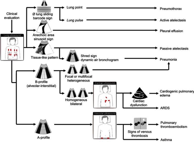
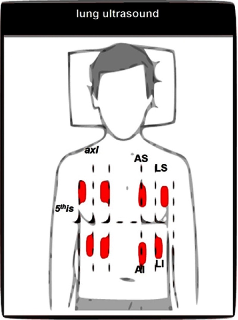
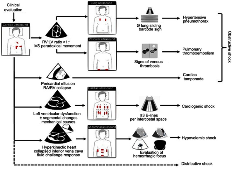
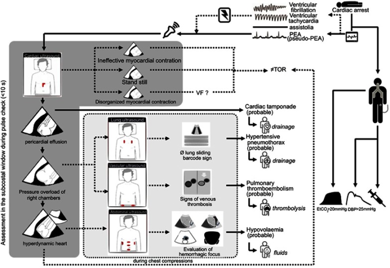
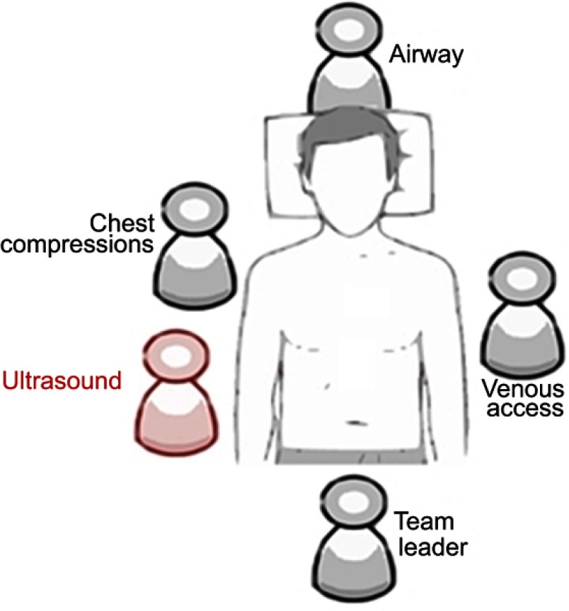
Ultrasound technology is an essential tool in the management of critically ill patients. Point-of-care ultrasonography (POCUS) enables data collection from different anatomic areas to achieve the most probable diagnosis and administer the right therapy at the right time. Despite the increasing utilization of POCUS, there is still a lack of standards to establish how to use different bedside ultrasound protocols, and it is imperative to develop a unifying protocol. Thus, the aim of this paper is to establish a new systematized approach that can be adopted by all physicians to implement POCUS for critically ill patient management. To achieve this, we propose a new systematized approach-Global Ultrasound Check for the Critically Ill (GUCCI)-that integrates multiple protocols. This protocol is organized based on three syndromes (acute respiratory failure, shock, and cardiac arrest) and includes ultrasound-guided procedures.
Keywords: cardiac arrest; echocardiography; intensive care; interventional ultrasonography; respiratory failure; shock; ultrasonography.
Conflict of interest statement
The authors declare that they have no conflict of interest in this work.
Figures


Similar articles
-
Global Ultrasound Check for the Critically Lll (GUCCI)-a New Systematized Protocol Unifying Point-of-Care Ultrasound in Critically Ill Patients Based on Clinical Presentation [Retraction].Open Access Emerg Med. 2022 Jun 23;14:273-274. doi: 10.2147/OAEM.S378669. eCollection 2022. Open Access Emerg Med. 2022. PMID: 35770140 Free PMC article.
-
Point-of-care ultrasound for non-vascular invasive procedures in critically ill neonates and children: current status and future perspectives.Eur J Pediatr. 2024 Mar;183(3):1037-1045. doi: 10.1007/s00431-023-05372-8. Epub 2023 Dec 12. Eur J Pediatr. 2024. PMID: 38085280 Review.
-
Emerging concepts in heart failure management and treatment: focus on point-of-care ultrasound in cardiogenic shock.Drugs Context. 2023 Jan 4;12:2022-5-8. doi: 10.7573/dic.2022-5-8. eCollection 2023. Drugs Context. 2023. PMID: 36660015 Free PMC article. Review.
-
[A Chinese consensus statement on the clinical application of transesophageal echocardiography for critical care (2019)].Zhonghua Nei Ke Za Zhi. 2019 Dec 1;58(12):869-882. doi: 10.3760/cma.j.issn.0578-1426.2019.12.002. Zhonghua Nei Ke Za Zhi. 2019. PMID: 31775449 Chinese.
-
"Playing it SAFE in the NICU" SAFE-R: a targeted diagnostic ultrasound protocol for the suddenly decompensating infant in the NICU.Eur J Pediatr. 2022 Jan;181(1):393-398. doi: 10.1007/s00431-021-04186-w. Epub 2021 Jul 5. Eur J Pediatr. 2022. PMID: 34223967 Free PMC article. Review.
Cited by
-
Thirteen dogs and a cat with ultrasonographically detected gallbladder wall edema associated with cardiac disease.J Vet Intern Med. 2021 May;35(3):1342-1346. doi: 10.1111/jvim.16117. Epub 2021 Apr 7. J Vet Intern Med. 2021. PMID: 33826214 Free PMC article.
-
Point-of-care ultrasound for critically-ill patients: A mini-review of key diagnostic features and protocols.World J Crit Care Med. 2022 Mar 9;11(2):70-84. doi: 10.5492/wjccm.v11.i2.70. eCollection 2022 Mar 9. World J Crit Care Med. 2022. PMID: 35433316 Free PMC article. Review.
-
Accuracy of echocardiography and ultrasound protocol to identify shock etiology in emergency department.BMC Emerg Med. 2022 Jun 30;22(1):117. doi: 10.1186/s12873-022-00678-6. BMC Emerg Med. 2022. PMID: 35768775 Free PMC article.
-
Lung Ultrasound Score Predicts the Extravascular Lung Water Content in Low-Birth-Weight Neonates with Patent Ductus Arteriosus.Med Sci Monit. 2020 Jun 15;26:e921671. doi: 10.12659/MSM.921671. Med Sci Monit. 2020. PMID: 32538377 Free PMC article.
-
Usefulness of Multi-Organ Point-of-Care Ultrasound as a Complement to the Decision-Making Process in Internal Medicine.J Clin Med. 2022 Apr 18;11(8):2256. doi: 10.3390/jcm11082256. J Clin Med. 2022. PMID: 35456356 Free PMC article.
References
-
- Nicoll CD, Mark LC. Diagnostic testing & medical decision making In: Papadakis MA, McPhee SJ, Rabow MW., editors. Current Medical Diagnosis & Treatment. New York, NY: McGraw-Hill Education; 2017. Available from: http://accessmedicine.mhmedical.com/content.aspx?aid=1145380368
Publication types
LinkOut - more resources
Full Text Sources
