Review
. 2019 Jul 25:8:F1000 Faculty Rev-1191.
doi: 10.12688/f1000research.18903.1. eCollection 2019.

Developmental synapse remodeling in the cerebellum and visual thalamus

Affiliations
Review

Developmental synapse remodeling in the cerebellum and visual thalamus

Masanobu Kano et al. F1000Res. .

Abstract

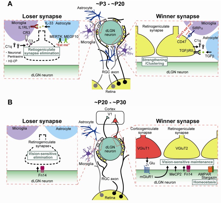
Functional neural circuits of mature animals are shaped during postnatal development by eliminating early-formed redundant synapses and strengthening of necessary connections. In the nervous system of newborn animals, redundant synapses are only transient features of the circuit. During subsequent postnatal development, some synapses are strengthened whereas other redundant connections are weakened and eventually eliminated. In this review, we introduce recent studies on the mechanisms of developmental remodeling of climbing fiber-to-Purkinje cell synapses in the cerebellum and synapses from the retina to neurons in the dorsal lateral geniculate nucleus of the visual thalamus (retinogeniculate synapses). These are the two representative models of developmental synapse remodeling in the brain and they share basic principles, including dependency on neural activity. However, recent studies have disclosed that, in several respects, the two models use different molecules and strategies to establish mature synaptic connectivity. We describe similarities and differences between the two models and discuss remaining issues to be tackled in the future in order to understand the general schemes of developmental synapse remodeling.

Keywords: Purkinje cell; cerebellum; climbing fiber; development; dorsal lateral geniculate nucleus; retinal ganglion cell; synapse remodeling.


Conflict of interest statement

No competing interests were disclosed.Competing interests: Roy V. Sillitoe has collaborated with Masanobu Kano on a 2016 consensus paper. Competing interests: Richard Hawkes has collaborated with Masanobu Kano on a 2016 consensus paper.

Figures

Figure 1.
Figure 1.. Remodeling of CF and PF synaptic connections onto PCs during postnatal cerebellar development.
(Upper panel) Schematics depicting developmental changes in CF and PF synaptic connections to PCs at ~P3, ~P7, ~P15, and ~P30. (Lower panel) Key events related to postnatal development of CF-to-PC and PF-to-PC synapses from birth to ~P30. BC, basket cell; CF, climbing fiber; GrC, granule cell; PC, Purkinje cell; PF, parallel fiber; SC, stellate cell.
Figure 2.
Figure 2.. Mechanisms for neural activity-mediated remodeling of CF-to-PC synapses during postnatal cerebellar development.
( A) Ca 2+ influx through P/Q-VDCC into PCs triggers strengthening of a single CF at ~P3 to ~P7 (functional differentiation). ( B) Ca 2+ influx through P/Q-VDCC promotes translocation of the single strong CF to the PC dendrite and at the same time eliminates CF synapses from the PC soma at ~P7 to ~P11 (early phase of CF elimination). GABAergic inhibition from BC to PC inhibits Ca 2+ influx and thereby regulates elimination of somatic CFs from ~P10. ( C) mGluR1 to PKCγ signaling and Arc activated by Ca 2+ influx through P/Q-VDCC promotes elimination of somatic CF synapses at ~P12 to ~P17 (late phase of CF elimination). ( D) mGluR1 and its downstream signaling in PCs promote elimination of PF synapses from proximal portions of PC dendrites from ~P15 to ~P30 (PF synapse elimination). BC, basket cell; CF, climbing fiber; mGluR1, metabotropic glutamate receptor subtype 1; PC, Purkinje cell; PF, parallel fiber; PKCγ, protein kinase Cγ; VDCC, voltage-dependent Ca 2+ channel.
Figure 3.
Figure 3.. Molecular and cellular mechanisms for transcellular interaction underlying elimination of loser CFs and strengthening/maintenance of winner CFs.
Bai3, brain-specific angiogenesis inhibitor 3; BC, basket cell; BDNF, brain-derived neurotrophic factor; BG, Bergmann glia; CF, climbing fiber; GLAST, L-glutamate/L-aspartate transporter; ItgB1, integrin B1; mGluR1, metabotropic glutamate receptor subtype 1; PC, Purkinje cell; PF, parallel fiber; PlxnA4, Plexin A4; PlxnC1, Plexin C1; Sema3A, Semaphorin 3A; Sema7A, Semaphorin 7A; Sort1, Sortilin 1; TrkB, tropomyosin receptor kinase B.
Figure 4.
Figure 4.. Remodeling of retinogeniculate synapses during postnatal development of the dorsal lateral geniculate nucleus (dLGN).
(Upper panel) Schematics depicting developmental changes in retinogeniculate synaptic connections to PCs at ~P5, ~P12, ~P20, ~P30, and ~P60. (Lower panel) Key events related to remodeling of retinogeniculate synaptic connections during postnatal development of the dLGN. RGC, retinal ganglion cell.
Figure 5.
Figure 5.. Molecular and cellular mechanisms underlying developmental remodeling of retinogeniculate synapses.
Molecular and cellular mechanisms underlying retinogeniculate synapse remodeling ( A) from ~P3 to ~P20 (eye-specific segregation and synapse elimination) and ( B) from ~P20 to ~P30 (vision-dependent synapse maintenance). dLGN, dorsal lateral geniculate nucleus; IL, interleukin; MeCP2, methyl-CpG-binding protein 2; mGluR1, metabotropic glutamate receptor subtype 1; RGC, retinal ganglion cell; SIRPα, signal regulatory protein alpha; TGF-β, transforming growth factor-beta; V1, primary visual cortex.

Similar articles

Cited by

References

    1. Hashimoto K, Kano M: Synapse elimination in the developing cerebellum. Cell Mol Life Sci. 2013;70(24):4667–80. 10.1007/s00018-013-1405-2 - DOI - PMC - PubMed
    1. Kano M, Hashimoto K: Synapse elimination in the central nervous system. Curr Opin Neurobiol. 2009;19(2):154–61. 10.1016/j.conb.2009.05.002 - DOI - PubMed
    1. Kano M, Watanabe T, Uesaka N, et al. : Multiple phases of climbing fiber synapse elimination in the developing cerebellum. Cerebellum. 2018;17(6):722–34. 10.1007/s12311-018-0964-z - DOI - PubMed
    1. Katz LC, Shatz CJ: Synaptic activity and the construction of cortical circuits. Science. 1996;274(5290):1133–8. 10.1126/science.274.5290.1133 - DOI - PubMed
    1. Lichtman JW, Colman H: Synapse elimination and indelible memory. Neuron. 2000;25(2):269–78. 10.1016/S0896-6273(00)80893-4 - DOI - PubMed

Publication types

LinkOut - more resources