. 2019 Jul 12:6:692-702.
doi: 10.1016/j.toxrep.2019.06.020. eCollection 2019.

Chemical profile and in vivo toxicity evaluation of unripe Citrus aurantifolia essential oil

Affiliations

Chemical profile and in vivo toxicity evaluation of unripe Citrus aurantifolia essential oil

Christian K Adokoh et al. Toxicol Rep. .

Abstract

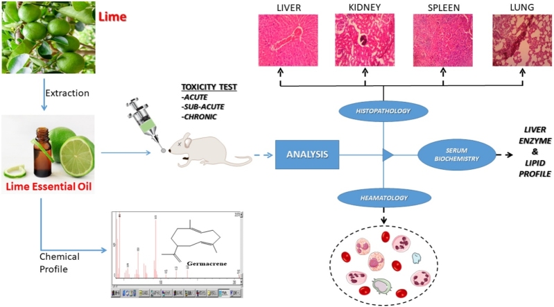
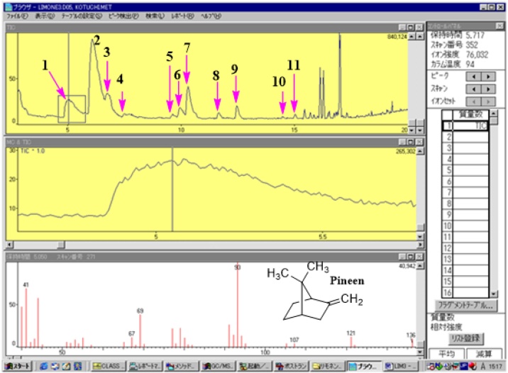
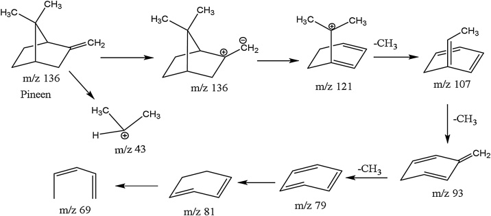
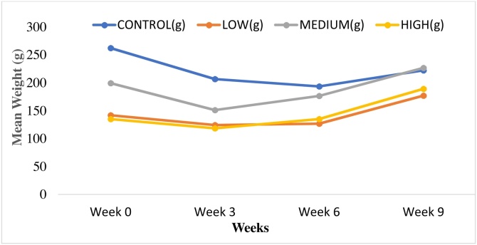
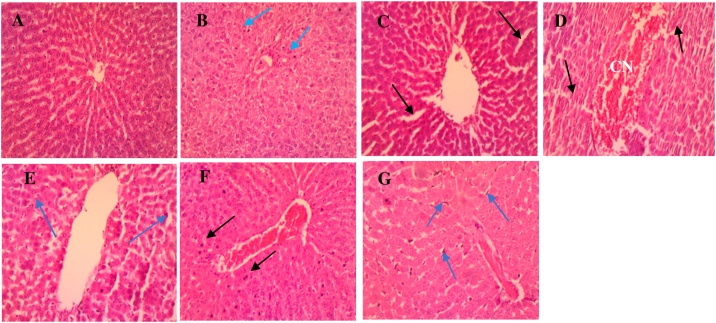
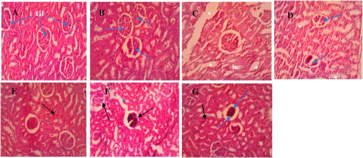
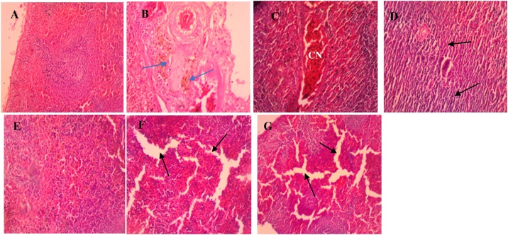
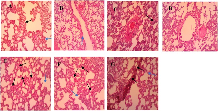
Citrus aurantifolia (Christm.) Swingle (syn. C. MEDICA var. ACIDA Brandis) (family: Rutaceae) essential oil is one of the cheapest oils found in local markets. Although, it is generally accepted as non-toxic to vital organs and cells, majority of people are cynical about it usage. Herein, the present study reports the chemical composition and in vivo oral toxicity study of unripe C. aurantifolia essential oil found in Ghana. The toxicity of C. aurantifolia essential oil extract was investigated via oral administration using two methods: The acute toxicity single dose study (SDS) and the repeated dose method. The oil exhibited no acute toxicity but in the sub-chronic studies, the effects was dose and time-dependent. Chemical profile investigation of the oil showed 9 constituent of phytochemicals (Germacrene isomers (61.2%), Pineen (14%), Linalool dimmer (2.9%), Bornane (11%), Citral (2.9%), Anethole (1.5%), Anisole (1.1%), Safrole (0.3%) and Demitol (0.6%)). Histopathological studies revealed conditions such as necrosis, edema and inflammatory reaction in the liver, spleen and kidneys. Marginal upsurge of biochemical parameters above normal and elevated levels of lymphocytes (35.20-46.40 g/dL) demonstrated mild toxicity among the 100 mg/kg and 500 mg/kg dose groups at the sub-chronic stage. Low levels of hemoglobin (13.60 to 12.70 g/dL), MCV (34.20-24.0 fL), MCH (40.20-36.40 g/dL) along with high levels of liver enzymes confirmed the mild toxicity of the oil at sub-chronic stage. These results demonstrate that, despite consideration of lime essential oil as safe, it can have mild hematotoxic, nephrotoxic and hepatotoxic effects.

Keywords: Biochemical analysis; Citrus aurantifolia; Hematological; Hepatotoxicity; Histopathological; In vivo toxicity; Nephrotoxicity.


Figures

None
Graphical abstract
Fig. 1
Fig. 1
GC/MS of unripe lime essential oil showing MS fragment of pinene.
Scheme 1
Scheme 1
Fragmentation pattern of beta pinene.
Fig. 2
Fig. 2
A graph of mean weights (g) against weeks for the control, low, medium and high dose groups.
Fig. 3
Fig. 3
Photomicrographs of liver sections for the normal control (A), sub-acute (B–D) and sub-chronic (E–G) stage. (A) Control group: shows hepatocytes with normal cytoarchitecture devoid of injuries. (B) Low dose: shows very few pyknotic nuclei (blue arrow) of hepatocytes. (C) Medium dose: shows sinusoidal dilatation accompanied by mild oedema (black arrows), with very mild leukocytosis. (D) High dose: shows congestion (CN) within the central vein along with distortion of architecture of the tissue (black arrow) and mild leukocytosis. (E) Low dose: shows marked leukocyte infiltration (arrow heads). (F) Medium dose: CN of central vein and focal pyknotic nuclei (black arrows) present. (G) High dose: mild dilation of sinusoids, severe Kupffer cell activation (blue arrows) and CN within sinusoids and central vein (H & E × 100).
Fig. 4
Fig. 4
Photomicrographs of kidney sections for the normal control (A), sub-acute (B–D) and sub-chronic (E–G) stage. (A) Control: normal glomeruli (Gl) and tubules showing no injuries. (B) Low dose: shows normal histology of Gl (blue arrows) as compared to the normal group. (C) Medium dose: shows oedema (white asterisks) and hydropic changes (hc). (D) High dose: shows oedema (asterisks) and Gl necrosis (blue arrows). Also, distortion of Gl structure and the tubules, are indicative of tissue damage (E) Low dose: Normal histology as compared to the normal group, with presence of congested vessels (black arrow). (F) Medium dose: marked glomerular necrosis (black arrows) and oedema (blue asterisk) marked by dilation of the tubules. (G) High dose: shows few pyknotic cells (black arrow) of PCT and DCT, increased Bowman’s space (blue arrow head) caused by Gl atrophy and necrosis (blue arrow) (H & E × 100).
Fig. 5
Fig. 5
Photomicrographs of spleen sections for the normal control (A), sub-acute (B–D) and sub-chronic (E–G) stage. (A) Control: normal tissue with the red and white pulps and the marginal zone separating them (B) Low dose: shows accumulation of hemosiderin (blue arrows) within the reticuloendothelial cells (blue arrows). (C) Medium dose: shows venous congestion (CN) with a little tissue distortion as compared to the control and low dose groups. (D) High dose: distortion of reticuloendothelial tissue and cytoarchitecture (black arrows) of spleen and atrophy of cells with marked spaces between adjacent immune cells. (E) Low dose: deposition of hemosiderin in the reticuloendothelial tissues. (F) Medium dose: shows sinusoidal dilatation and oedema (black arrows). (G) High dose: shows sinusoidal dilatation evident as oedema (black arrows), more severe than what is observed in the medium dose group (H & E × 40).
Fig. 6
Fig. 6
Photomicrographs of lungs sections for the normal control (A), sub-acute (B–D) and sub-chronic (E–G) stage. (A) Control: shows normal alveolar duct with its walls consisting of adjacent alveoli (blue arrow) separated by interalveolar septa (black arrow). (B) Low dose: has normal histology as compared to the normal control group. (C) Medium dose: mild leukocytosis (blue arrow) with thickened aveoli septa (black arrow). (D) High dose: shows the presence of few inflammatory cells. (E) Low dose: slightly thickened aveoli septa (blue arrow), accompanied by focal inter-aveoli congestion (black arrows) (F) Medium dose: moderately thickened aveoli septa (blue arrow) accompanied by mild inter-aveoli congestion (black arrows) (G) High dose: shows marked leukocytic infiltrate within sections (H&E × 100).

References

    1. Narang N., Jiraungkoorskul W. Anticancer activity of key lime. Citrus aurantifolia Pharmacogn. Rev. 2016;10:118–122. - PMC - PubMed
    1. Ivine F.R. Oxford University Press; Oxford: 1961. Woody Plants of Ghana; p. 493.
    1. Perry N., Perry E. Aromatherapy in the management of psychiatric disorders clinical and neuropharmacological perspectives. CNS Drugs. 2006;20:257–280. - PubMed
    1. Jimbo D., Kimura Y., Taniguchi M., Inoue M., Urakami K. Effect of aromatherapy on patients with Alzheimer’s disease. Psychogeriatrics. 2009;9:173–179. - PubMed
    1. Smith C.A., Collins C.T., Crowther C.A. Aromatherapy for pain management in labour. Cochrane Database Syst. Rev. 2011 - PubMed