Case Reports
. 2019 Jul 12;9(3):e2019104.
doi: 10.4322/acr.2019.104. eCollection 2019 Jul-Sep.

Uterine cavity embryonal rhabdomyosarcoma

Affiliations
Case Reports

Uterine cavity embryonal rhabdomyosarcoma

Marina Gomes Pereira Sardinha et al. Autops Case Rep. .

Abstract

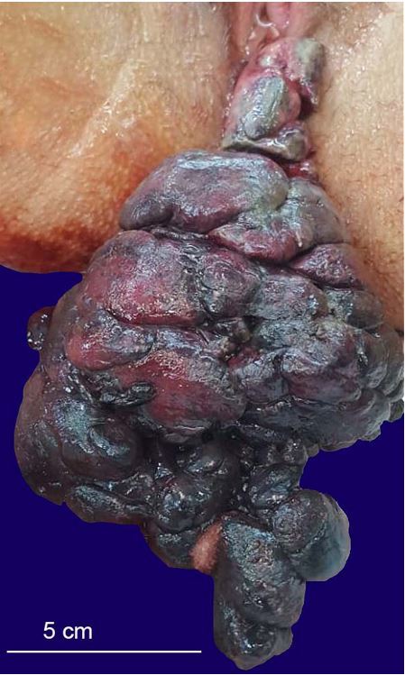
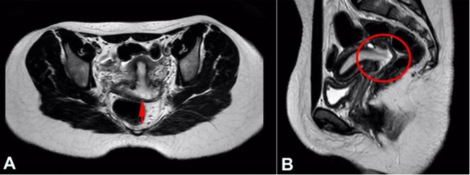
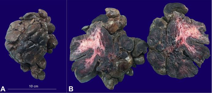
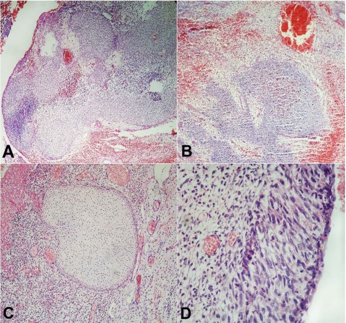
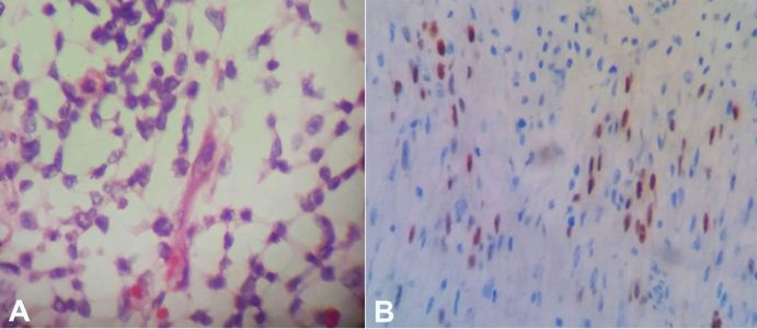
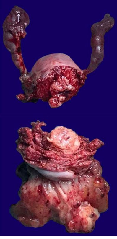
Rhabdomyosarcoma (RMS) is a rare solid tumor in childhood and adolescence. The higher incidence is predominant during the first two decades of life. According to the Intergroup RMS Study Group, the embryonal RMS (ERMS), botryoidal variant, constitutes a histological subtype characterized as a "grape-like" lesion of 2.0 cm to 9.5 cm. The treatment involves chemotherapy, surgery, and/or radiotherapy. We present the case of a 14-year-old female patient diagnosed with ERMS, botryoidal variant, which originated in the uterine cervix with vaginal externalization. The initial therapeutic approach comprised an initial prolapsed mass excision followed by Wertheim-Meigs surgery due to the tumor extension. No consensual protocol to ERMS treatment is found in the medical literature; however, a combined approach seems to offer a better result. The postoperative time period was uneventful and the patient followed an adjuvant therapy with vincristine, d-actinomycin, and cyclophosphamide. A comprehensive evaluation of the therapeutic options preserving the reproductive function-unfortunately not always possible-is part of a multi-disciplined care team concerning the pediatric patients.

Keywords: Cervix Uteri; Rhabdomyosarcoma, Embryonal; Uterine Cervical Neoplasms.


Conflict of interest statement

Conflict of interest: None

Figures

Figure 1
Figure 1. Large, foul-smelling, lobed-shaped vaginal exophytic lesion of 14 × 10 × 8 cm attached to the uterine cervix by a pedicle.
Figure 2
Figure 2. A – Axial cut T2 and its correspondence in sagittal cut T2 (B) of the magnetic resonance imaging of the pelvis showing a uterus with normal dimensions, diffuse thickening of the endometrium (arrow and circled area)—more significant in the posterior region of the uterine cervix, and irregular contour of the adjacent myometrium, possibly corresponding to neoplastic infiltration.
Figure 3
Figure 3. A – Surgical excision specimen was a blackish bunch-of-grapes-like mass of 14.0 × 10.0 × 8.0 cm weighing 0.468 kg; B – Hardened tissue with necrosis and a fleshy pink center.
Figure 4
Figure 4. Photomicrographs of the undifferentiated neoplasia: A – Subepithelial hypercellular areas (H&E, 40X); B – Extensive necro hemorrhagic component of neoplasia (H&E, 40X); C – Foci of cartilaginous metaplasia (H&E,40X); D – Prevalence of small, round, blue cells, characterizing a probable neoplasm of embryonal cells (H&E, 100X).
Figure 5
Figure 5. Photomicrographs of the tumor. A – Rare elongated cells with eosinophilic cytoplasm, suggesting rhabdomyoblastic differentiation (H&E, 400X); B – Immunohistochemistry positivity for myogenin.
Figure 6
Figure 6. Gross view of the surgical specimen after the Wertheim-Meigs surgery showing residual “grape-like” gelatinous tumor mass within the uterine cavity (edited picture).

References

    1. Raney RB, Maurer HM, Anderson JR, et al. . The Intergroup Rhabdomyosarcoma Study Group (IRSG): major lessons from the IRS-I through IRS-IV studies as background for the current IRS-V treatment protocols. Sarcoma. 2001;5(1):9-15. 10.1080/13577140120048890. - DOI - PMC - PubMed
    1. Chintagumpala MM. Soft-tissue sarcomas In: McMillan JA, Feigin RD, DeAngelis CD, Jones MD Jr, editors. Oski’s pediatrics: principles and practice. 4th ed. Philadelphia: Williams & Wilkins; 2006. p. 1781-6.
    1. Maurer HM. The Intergroup Rhabdomyosarcoma study: update. Natl Cancer Inst Monogr. 1981;56(56):61-8. - PubMed
    1. Semczuk A, Baranowski W, Berbeć H, Marzec B, Skomra D, Miturski R. Analysis of p53 and K-ras genes and their proteins in a sarcoma botryoides of the uterine cervix. Eur J Gynaecol Oncol. 1999;20(4):311-4. - PubMed
    1. Behtash N, Mousavi A, Tehranian A, Khanafshar N, Hanjani P. Embryonal rhabdomyosarcoma of the uterine cervix: case report and review of the literature. Gynecol Oncol. 2003;91(2):452-5. 10.1016/S0090-8258(03)00539-0. - DOI - PubMed

Publication types