. 2020 Feb;42(2):193-199.
doi: 10.1007/s00276-019-02298-5. Epub 2019 Aug 1.

Anatomical variations of the dentate gyrus in normal adult brain

Affiliations

Anatomical variations of the dentate gyrus in normal adult brain

Robert Haładaj. Surg Radiol Anat. 2020 Feb.

Abstract

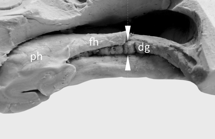
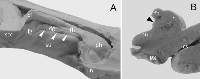
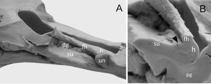
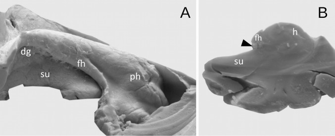
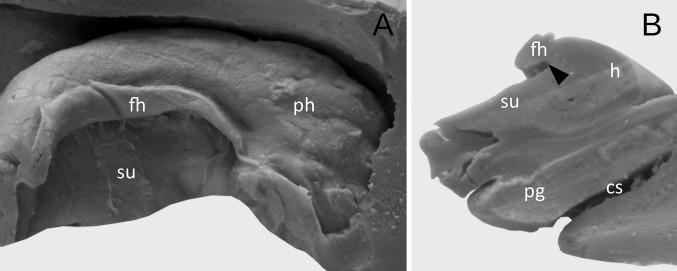
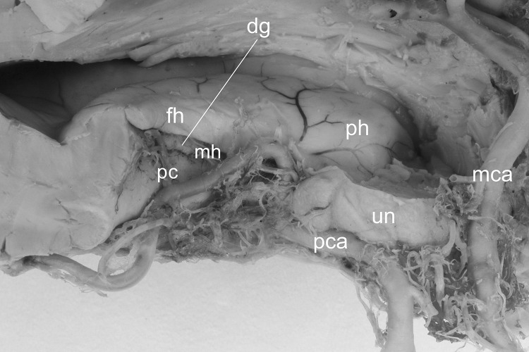
Recent scientific papers indicate the clinical significance of the dentate gyrus. However, a detailed knowledge of the anatomical variations of this structure in normal adult brain is still lacking. An understanding of the variable morphology of the dentate gyrus may be important for diagnostic neuroimaging. Thus, the purpose of this macroscopic cadaveric study was to describe the anatomical variations of the dentate gyrus. Forty formalin-fixed human cerebral hemispheres, obtained from bodies of donors without the history of neuropathological diseases, were included in the study. The dentate gyrus was classified as well-developed, when it protruded completely from under the fimbria of the hippocampus. The gyrus was classified as underdeveloped, when it was covered by the fimbria of the hippocampus (but clearly visible at the coronal section of the hippocampal formation), while the hypoplastic gyrus was not visible macroscopically under the fimbria of the hippocampus. The well-developed type was observed in 27 cases (67.5%). The thickness of well-developed type of the dentate gyrus, measured between the fimbriodentate sulcus and hippocampal sulcus, varied from 2.74 to 5.21 mm (mean = 3.67 mm, median = 5.54 mm, SD 0.65 mm). In the next nine cases (22.5%), the dentate gyrus was underdeveloped. The thickness of underdeveloped type of the dentate gyrus varied from 1.75 to 2.37 mm (mean = 2.02 mm, median = 2.16 mm, SD 0.33 mm). In the remaining four cases (10%), the dentate gyrus was hypoplastic and could not be distinguished macroscopically. In all injected hemispheres, arterial supply of the dentate gyrus was provided by the branches of the posterior cerebral artery. Awareness of normal variations of the dentate gyrus may allow for better correlation of anatomical knowledge with radiological data and for use this knowledge to describe abnormal conditions.

Keywords: Cerebral cortex; Dentate gyrus; Hippocampal arteries; Hippocampal formation; Human brain; Temporal lobe.


Conflict of interest statement

There is no conflict of interest to declare.

Figures

Fig. 1
Fig. 1
Assessment of the dentate gyrus morphology. The well-developed margo denticulatus of the dentate gyrus (dg) was visualized on the medial side of the fimbria of the hippocampus (fh). The tooth-shaped part of the dentate gyrus is clearly visible. For comparison purposes, horizontal incisions through each hippocampal formation were performed with the aid of a brain knife, in the half-length of the body of the hippocampus (place of the incision was marked by white arrowheads). At this level the thickness of the margo denticulatus was also measured, between the fimbriodentate sulcus and hippocampal sulcus. ph pes of the hippocampus
Fig. 2
Fig. 2
The appearance of the well-developed dentate gyrus. a Medial view. The dentate gyrus (dg) was classified as well-developed, when the margo denticulatus protruded completely from under the fimbria of the hippocampus (fh). In those cases, the hippocampal sulcus (marked by white arrowheads) was well-developed and clearly visible along all length of the margo denticulatus. b The coronal section through the hippocampal formation showing the well-developed dentate gyrus (marked by black arrowhead). cf crus of fornix, cs collateral sulcus, fg fasciolar gyrus, h hippocampus proper (Ammon’s horn), pg parahippocampal gyrus, ph pes of hippocampus, scc splenium of the corpus callosum, su subiculum of the parahippocampal gyrus, un uncus
Fig. 3
Fig. 3
The appearance of the well-developed dentate gyrus with irregular prominences and less marked hippocampal sulcus. a Medial view. b The coronal section through the hippocampal formation showing the dentate gyrus (marked by black arrowhead). h hippocampus proper (Ammon’s horn), dg margo denticulatus of the dentate gyrus, fh fimbria of the hippocampus, pg parahippocampal gyrus, ph pes of hippocampus, su subiculum of the parahippocampal gyrus
Fig. 4
Fig. 4
The appearance of the underdeveloped dentate gyrus. a Medial view. The dentate gyrus was classified as underdeveloped, when the margo denticulatus (dg) was covered by the fimbria of the hippocampus (fh). b The coronal section through the hippocampal formation showing the underdeveloped margo denticulatus (marked by black arrowhead). h hippocampus proper (Ammon’s horn), ph pes of hippocampus, su subiculum of the parahippocampal gyrus
Fig. 5
Fig. 5
Hypoplasia of the dentate gyrus. a Medial view. The dentate gyrus was classified as underdeveloped, when the margo denticulatus was not visible macroscopically under the fimbria of the hippocampus (fh). b The coronal section through the hippocampal formation showing the hypoplasia of the dentate gyrus (black arrowhead shows the place with absent or vestigial margo denticulatus of dentate gyrus). cs collateral sulcus, h hippocampus proper (Ammon’s horn), pg parahippocampal gyrus, ph pes of hippocampus, su subiculum of the parahippocampal gyrus
Fig. 6
Fig. 6
Arterial supply of the dentate gyrus. Arterial supply of the dentate gyrus is provided by the branches of the posterior cerebral artery (pca), namely by one of the posterior choroidal arteries (pc) and by the middle hippocampal artery (mh). dg margo denticulatus of the dentate gyrus, fh fimbria of the hippocampus, h hippocampus proper (Ammon’s horn), mca middle cerebral artery, ph pes of hippocampus, un uncus

References

    1. Amaral DG, Lavenex P. Hippocampal neuroanatomy. In: Andersen P, Morris R, Amaral D, Bliss T, O’Keefe J, editors. The hippocampus book. New York: Oxford University Press; 2007. p. 872.
    1. Amaral DG, Scharfman HE, Lavenex P. The dentate gyrus: fundamental neuroanatomical organization (dentate gyrus for dummies) Prog Brain Res. 2007;163:3–22. doi: 10.1016/S0079-6123(07)63001-5. - DOI - PMC - PubMed
    1. Boldrini M, Galfalvy H, Dwork AJ, Rosoklija GB, Trencevska-Ivanovska I, Pavlovski G, Hen R, Arango V, Mann JJ. Resilience is associated with larger dentate gyrus, while suicide decedents with major depressive disorder have fewer granule neurons. Biol Psychiatry. 2019 doi: 10.1016/j.biopsych.2018.12.022. - DOI - PMC - PubMed
    1. Clark AW, Sarnat HB. Bilateral agenesis of the hippocampal dentate gyrus in a neurologically normal adult. Can J Neurol Sci. 2006;33:296–301. doi: 10.1017/S0317167100005163. - DOI - PubMed
    1. Daugherty AM, Flinn R, Ofen N. Hippocampal CA3-dentate gyrus volume uniquely linked to improvement in associative memory from childhood to adulthood. Neuroimage. 2017;153:75–85. doi: 10.1016/j.neuroimage.2017.03.047. - DOI - PMC - PubMed

LinkOut - more resources