. 2019 Aug 1;5(1):11.
doi: 10.1186/s41205-019-0048-9.

Advanced 3D printed model of middle cerebral artery aneurysms for neurosurgery simulation

Affiliations

Advanced 3D printed model of middle cerebral artery aneurysms for neurosurgery simulation

Ruth G Nagassa et al. 3D Print Med. .

Abstract

Background: Neurosurgical residents are finding it more difficult to obtain experience as the primary operator in aneurysm surgery. The present study aimed to replicate patient-derived cranial anatomy, pathology and human tissue properties relevant to cerebral aneurysm intervention through 3D printing and 3D print-driven casting techniques. The final simulator was designed to provide accurate simulation of a human head with a middle cerebral artery (MCA) aneurysm.

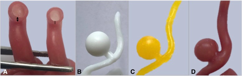
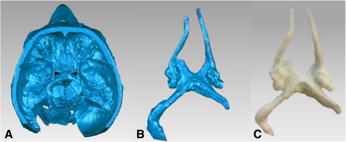
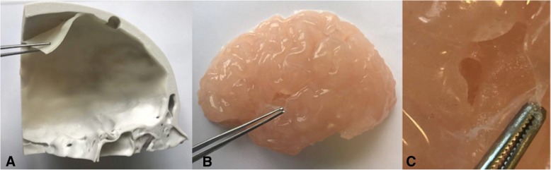
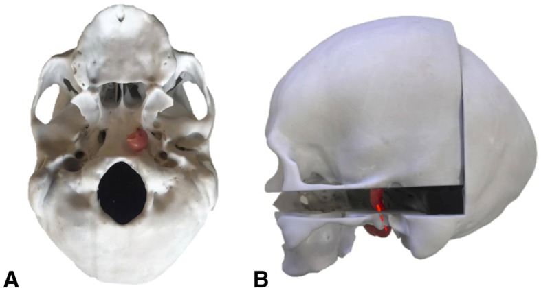
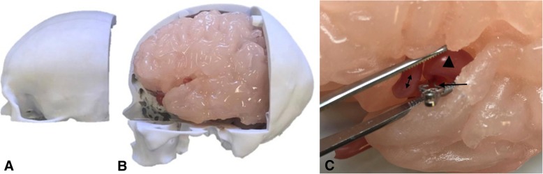
Methods: This study utilized living human and cadaver-derived medical imaging data including CT angiography and MRI scans. Computer-aided design (CAD) models and pre-existing computational 3D models were also incorporated in the development of the simulator. The design was based on including anatomical components vital to the surgery of MCA aneurysms while focusing on reproducibility, adaptability and functionality of the simulator. Various methods of 3D printing were utilized for the direct development of anatomical replicas and moulds for casting components that optimized the bio-mimicry and mechanical properties of human tissues. Synthetic materials including various types of silicone and ballistics gelatin were cast in these moulds. A novel technique utilizing water-soluble wax and silicone was used to establish hollow patient-derived cerebrovascular models.

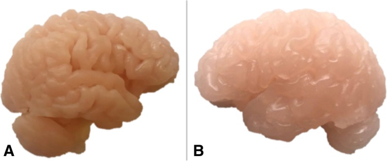
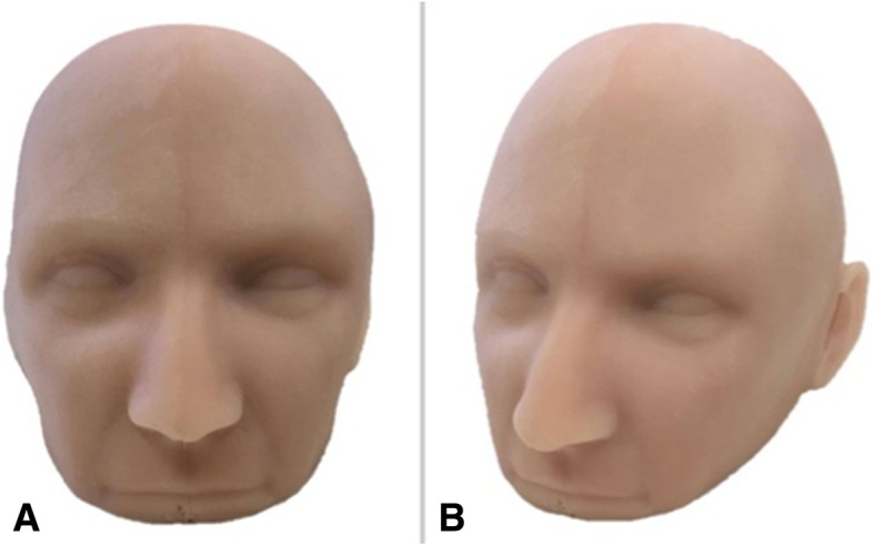
Results: A patient-derived 3D aneurysm model was constructed for a MCA aneurysm. Multiple cerebral aneurysm models, patient-derived and CAD, were replicated as hollow high-fidelity models. The final assembled simulator integrated six anatomical components relevant to the treatment of cerebral aneurysms of the Circle of Willis in the left cerebral hemisphere. These included models of the cerebral vasculature, cranial nerves, brain, meninges, skull and skin. The cerebral circulation was modeled through the patient-derived vasculature within the brain model. Linear and volumetric measurements of specific physical modular components were repeated, averaged and compared to the original 3D meshes generated from the medical imaging data. Calculation of the concordance correlation coefficient (ρc: 90.2%-99.0%) and percentage difference (≤0.4%) confirmed the accuracy of the models.

Conclusions: A multi-disciplinary approach involving 3D printing and casting techniques was used to successfully construct a multi-component cerebral aneurysm surgery simulator. Further study is planned to demonstrate the educational value of the proposed simulator for neurosurgery residents.

Keywords: 3D printing; Anatomical models; Aneurysm; Neurosurgical training; Simulation.


Conflict of interest statement

The authors declare that they have no competing interests.

Figures

Fig. 1
Fig. 1
Flowchart presents the workflow for simulator development. Initially, DICOM data was imported into segmentation software in which the region of interest was isolated. The 3D rendered mesh was processed. The generated .STL files were assigned to a 3D printer to print anatomical models and moulds. The moulds were used to cast soft-tissue models in various materials. Post-print processing involved the removal of 3D printing support materials and assembly of the six-component simulator
Fig. 2
Fig. 2
a Terminal branches of the patient-derived vasculature lumen (left MCA) in which two layers of silicone (line) produced a thinner, more accurate representation of vessel wall thickness than that of three layers (double sided arrow). b 3D print of CAD aneurysm model used to develop c a wax cast and d hollow silicone model
Fig. 3
Fig. 3
a Computational model of dissected specimen (MP1670) with the optic chiasm, optic nerve and olfactory tract (between arrows) that were isolated b as a separate mesh and c 3D printed through FDM technology to achieve a flexible 3D printed model
Fig. 4
Fig. 4
a Right cerebral hemisphere cast in Clear Ballistics gel 10% with firmer material properties to that of the b left cerebral hemisphere cast in Clear Ballistics medical gel #4
Fig. 5
Fig. 5
a Dura mater model pulled off the calvaria roof demonstrating the thickness and color consistency of the model achieved through silicone application. b Brain model (left hemisphere) with pia mater attached and dissected at the Sylvian fissure. c Amplified view demonstrating the thin transparent silicone layer consistent with the characteristic of real tissue
Fig. 6
Fig. 6
a Inferior view and b lateral view of the skull model with the vasculature model introduced through the carotid canal across the two skull components (dashed red line)
Fig. 7
Fig. 7
a Frontal view of the silicone skin model and b angled view permitting visualization of the left ear that serves as a guide for scalp incision during surgical intervention
Fig. 8
Fig. 8
a Removal of replaceable bone flap with adhered dura mater model on the interior surface, exposing b the left cerebral hemisphere with adhered pia mater model. c Retraction of the Sylvian fissure of the brain model and application of a surgical clip (arrow) at the aneurysm neck of the vasculature model. Visualization of the aneurysm dome (triangle) and the neighboring M2 branch (double-sided arrow) of the patient-derived aneurysm model

References

    1. Lai L, Morgan MK. The impact of changing intracranial aneurysm practice on the education of cerebrovascular neurosurgeons. J Clin Neurosci. 2012;19(1):81–84. doi: 10.1016/j.jocn.2011.07.008. - DOI - PubMed
    1. Rehder R, Abd-El-Barr M, Hooten K, Weinstock P, Madsen JR, Cohen AR. The role of simulation in neurosurgery. Childs Nerv Syst. 2016;32(1):43–54. doi: 10.1007/s00381-015-2923-z. - DOI - PubMed
    1. Alaraj AT, Charbel FP, Birk DP, Tobin MP, Luciano CP, Banerjee PP, et al. Role of cranial and spinal virtual and augmented reality simulation using immersive touch modules in neurosurgical training. Neurosurgery. 2013;72(1):115–123. doi: 10.1227/NEU.0b013e3182753093. - DOI - PMC - PubMed
    1. Shakir HJ, Shallwani H, Levy EI. Editorial: see one, do one, teach one? Paradigm shift with three-dimensional printing. Neurosurgery. 2017;80(1):3–5. - PubMed
    1. Alaraj A, Luciano C, Bailey D, Elsenousi A, Roitberg B, Bernardo A, et al. Virtual reality cerebral aneurysm clipping simulation with real-time haptic feedback. Neurosurgery. 2015;11(1):52–58. - PMC - PubMed

LinkOut - more resources