Review
. 2019 Jul;7(4):10.1128/microbiolspec.gpp3-0045-2018.
doi: 10.1128/microbiolspec.GPP3-0045-2018.

Molecular Mimicry, Autoimmunity, and Infection: The Cross-Reactive Antigens of Group A Streptococci and their Sequelae

Affiliations
Review

Molecular Mimicry, Autoimmunity, and Infection: The Cross-Reactive Antigens of Group A Streptococci and their Sequelae

Madeleine W Cunningham. Microbiol Spectr. 2019 Jul.

Abstract

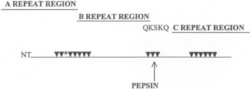
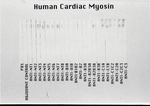
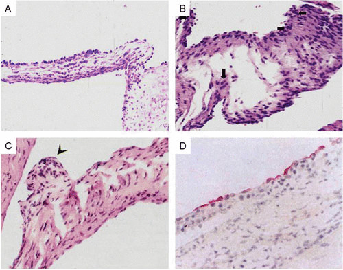
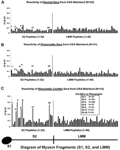
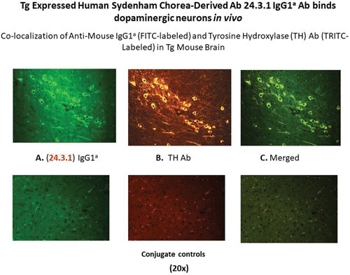
The group A streptococci are associated with a group of diseases affecting the heart, brain, and joints that are collectively referred to as acute rheumatic fever. The streptococcal immune-mediated sequelae, including acute rheumatic fever, are due to antibody and cellular immune responses that target antigens in the heart and brain as well as the group A streptococcal cross-reactive antigens as reviewed in this article. The pathogenesis of acute rheumatic fever, rheumatic heart disease, Sydenham chorea, and other autoimmune sequelae is related to autoantibodies that are characteristic of autoimmune diseases and result from the immune responses against group A streptococcal infection by the host. The sharing of host and streptococcal epitopes leads to molecular mimicry between the streptococcal and host antigens that are recognized by the autoantibodies during the host response. This article elaborates on the discoveries that led to a better understanding of the pathogenesis of disease and provides an overview of the history and the most current thought about the immune responses against the host and streptococcal cross-reactive antigens in group A streptococcal sequelae.


Figures

FIGURE 1
FIGURE 1
Reaction of mouse antistreptococcal mAb with a human tissue section of myocardium in a indirect immunofluorescence assay. Mouse IgM (20 μg/ml) was unreactive (not shown). mAbs were tested at 20 μg/ml. (Taken from reference with permission from the Journal of Immunology.)
FIGURE 2
FIGURE 2
Human and murine antistreptococcal/antimyosin mAbs were divided into three subsets based on their reactivity with myosin and N-acetyl-glucosamine, the dominant group A carbohydrate epitope, with DNA and the cell nucleus, a property found among murine mAbs, and reactivity with myosin and a family of alpha-helical coiled-coil molecules. (Taken from reference with permission from Indiana University School of Dentistry Press.)
FIGURE 3
FIGURE 3
Diagram illustrating the potential mechanism of antibody in the pathogenesis of rheumatic heart disease. Cross-reactive antibody is shown binding directly to endothelium (top diagram) or binding to basement membrane of the valve (bottom diagram) exposed due to shear stress or damage by antibody and complement. (Reproduced with permission from ASM press).
FIGURE 4
FIGURE 4
Reactivity of antistreptococcal/antimyosin mAb 3.B6 with normal human valve endothelium and myocardium. Formalin-fixed human mitral valve (A) and myocardium (B) were reacted with mAb 3.B6 at 10 μg/ml. mAb 3.B6 binding was detected using biotin-conjugated antihuman antibodies and alkaline phosphatase-labeled streptavidin followed by fast red substrate. Control sections did not react with human IgM at 10 μg/ml (C,D). (Reproduced with permission from Lippincott Williams and Wilkins).
FIGURE 5
FIGURE 5
Adhesion and extravasation of T lymphocytes into an ARF valve in valvulitis. (A,B) Extravasation of CD4+ lymphocytes (stained red; original magnification, 200× and 400×, respectively). (C) Extravasation of CD8+ lymphocytes (stained red) into the valve through the valvular endothelium (magnification, 200×). An IgG1 isotype control mAb (IgG1) did not react with the same valve (not shown). (With permission from the Journal of Infectious Diseases, Chicago Press).
FIGURE 6
FIGURE 6
Sequence alignment of streptococcal M6 protein and human cardiac tropomyosin in a region exhibiting significant homology. Lowercase letters a to g directly above the sequence designate the position of these amino acids within the seven-residue periodicity in both segments. Lowercase letters at the top of the figure designate identities at external locations in the heptad repeat. Double dots indicate identities, and single dots indicate conservative substitutions. Within this segment of the streptococcal M6 molecule, 31% homology is observed with tropomyosin. Since both molecules are alpha-helical coiled-coil proteins, they contain the seven-residue repeat pattern where positions a and d are usually hydrophobic. Similar homologies are seen between M proteins and myosin heavy chains and any of the three laminin chains. (Reproduced from reference with permission from the Journal of Immunology.)
FIGURE 7
FIGURE 7
Antimyosin antibody cross-reactive sites in M5 protein A, B, and C repeat regions of the molecule. Arrows point to sites determined to induce human cardiac myosin cross-reactive antibody (32). The asterisk (*) marks the location of peptide NT4 containing several repeats of an epitope in cardiac myosins that causes myocarditis in MRL/++ and BALB/c mouse strains (32, 116). Site QKSKQ is the epitope determined to react with antimyosin antibody in ARF sera (40). (Reproduced from Effects of Microbes on the Immune System with permission from Lippincott Williams and Wilkins.)
FIGURE 8
FIGURE 8
Reactivity of the antistreptococcal M5 peptide sera in a Western immunoblot of human cardiac myosin. The 200-kDa protein band of the purified human cardiac myosin, shown in the stained portion of the Western blot, reacted most strongly with antipeptide sera from mice immunized with the M5 peptides NT3-7, B2B3B, C1A, C1B, C2C3, and C3. Sera were tested at a 1:1000 dilution. A control antimyosin mAb CCM-52 (a gift from William Clark, Cardiovascular Research Institute, Michael Reese Hospital and Medical Center, Chicago, IL) reacted with the 200-kDa band present in our purified preparation of human cardiac myosin heavy chain. Purification of the human cardiac myosin heavy chain to homogeneity was previously described by Dell et al. (204). The Western blot confirms the data seen in the enzyme-linked immunosorbent assay with human cardiac myosin. (Reproduced from reference with permission from ASM Press.)
FIGURE 9
FIGURE 9
Sequence homology between human cardiac myosin and peptide NT4 that causes myocarditis (32, 119). The homologous sequence repeats four times in the streptococcal M5 protein and in NT4 and once in cardiac myosin. Repeated sequences in M proteins that mimic cardiac myosin may be important in inducing inflammatory heart disease. (Adapted from reference with permission from the Journal of Immunology.)
FIGURE 10
FIGURE 10
Diagram representing the proposed immunopathogenesis of poststreptococcal rheumatic heart disease. Initially, B and T cells are activated by specific streptococcal antigens and superantigens, leading to strong immune responses against streptococcal and host antigens. The development of pathogenic clones of B and T lymphocytes is important in the development of the disease. Initially, antibodies develop against the group A carbohydrate, which are cross-reactive with the valve surface, and glycoproteins such as laminin and bind to the valve surface endothelium (endocardium), leading to inflammation and upregulation of cell adhesion molecules such as VCAM-1 on activated surface endothelium of the valve. M protein-reactive T cells enter the valve through the surface endothelium by binding to cell adhesion molecules such as VCAM-1 and extravasate into the valve (48). The formation of scar tissue in the valve followed by neovascularization allows the disease to continue in the valve. The specificity of the T cells in blood (25) and T cells entering the valve have been shown to react to M protein (22, 97, 144). T cell subsets include Th1 (IFNγ) (145) in the pathogenesis of proinflammatory responses and the development of the scarred and fibrotic valve. IL-17A has also been associated with rheumatic heart disease in humans and animal models, suggesting that Th17 cells are involved in disease (101, 146, 149).
FIGURE 11
FIGURE 11
(A) Induction of valvulitis and cellular infiltration in hematoxylin- and eosin-stained heart valves from Lewis rats immunized with group A streptococcal M5 peptides NT1 to NT4/5 (AVTRGTINDPQRAKEALD amino acid [aa] residues 1 to 18; NT-2 KEALDKYELENHDLKTKN aa residues 14 to 31; NT-3 LKTKNEGLKTENEGLKTE aa residues 27 to 44; NT-4 GLKTENEGLKTENEGLKTE aa residues 40 to 58; NT-4/5 GLKTEKKEHEAENDKLK aa residues 54 to 70). (Taken from Kirvan et al. [8] with permission from the Journal of Translational Cardiology.) (B) Induction of valvulitis, edema, and cellular infiltration in hematoxylin- and eosin-stained heart valves from Lewis rats immunized with group A streptococcal M5 peptides NT1 to NT4/5 (AVTRGTINDPQRAKEALD aa residues 1 to 18; NT-2 KEALDKYELENHDLKTKN aa residues 14 to 31; NT-3 LKTKNEGLKTENEGLKTE aa residues 27 to 44; NT-4 GLKTENEGLKTENEGLKTE aa residues 40 to 58; NT-4/5 GLKTEKKEHEAENDKLK aa residues 54 to 70). (Taken from Kirvan et al. [8] with permission from the Journal of Translational Cardiology.) (C) Verrucous nodule observed on Lewis rat valve after immunization with group A streptococcal rM6 protein. (Taken from Quinn et al. [23] with permission from Infection and Immunity.) (D) VCAM-1 expressed on rheumatic valve. (Taken from Roberts et al. [48] with permission from the Journal of Infectious Diseases.)
FIGURE 12
FIGURE 12
Extravasation of CD4+ lymphocytes into valve above Aschoff’s body in the subendocardium of the left atrial appendage. Original magnification, 200×. (A) Stained with anti-CD4 Mab; (B) stained with antibody isotype control. (Taken from Roberts et al. [48]).
FIGURE 13
FIGURE 13
Reactivity of serum samples originating from the United States with human cardiac myosin peptides from the S2 and LMM regions in the enzyme-linked immunosorbent assay (1:100 serum dilution). (A) Mean reactivity of normal serum samples from control donors with no evidence of streptococcal infection or heart disease on the U.S. mainland against S2 and LMM peptides. These samples rarely reacted with any S2 or LMM peptide at an optical density of 0.2. (B) Mean reactivity of serum samples from patients with streptococcal pharyngitis in the United States against S2 and LMM peptides. These samples rarely reacted with any S2 or LMM peptide at an optical density of 0.2. (C) Serum immunoglobulin G from patients with rheumatic carditis in the United States. Serum samples from patients with rheumatic carditis from the United States reacted predominantly with peptides S2-1, S2-4, S2-5, S2-8, S2-9, S2-17, and S2-30, compared with the reactivity of serum samples from patients with pharyngitis in the United States against those same peptides (B). (C) Unadjusted Mann-Whitney P values for the comparison between carditis and pharyngitis on the United States mainland. The comparison for S2-4 is statistically significant on the basis of a two-sided alpha level adjusted to preserve the false-discovery rate at 5%. (Taken from Ellis et al. [24] with permission from the Journal of Infectious Diseases).
FIGURE 14
FIGURE 14
The human Sydenham chorea MAb 24.3.1 V gene expressed as the human V gene-mouse IgG1a constant region in transgenic (Tg) mice targets dopaminergic neurons. Chimeric Tg24.3.1 VH IgG1a antibody expressed in Tg mouse sera penetrated dopaminergic neurons in Tg mouse brain in vivo. Colocalization of Tg 24.3.1 IgG1a (anti-IgG1a Ab; left panel) and tyrosine hydroxylase antibody (anti-TH Ab; middle panel). TH is a marker for dopaminergic neurons. The left-hand panel shows IgG1a (labeled with fluorescein isothiocyanate [FITC]), the center panel shows TH Ab (labeled with tetramethylrhodamine isothiocyanate [TRITC]), and the right-hand panel shows a merged image (FITC-TRITC). Brain sections (basal ganglia) of VH24.3.1 Tg mouse (original magnification, 320×), showing (A) FITC-labeled anti-mouse IgG1a, (B) TRITC-labeled anti-TH Ab, and (C) the merged image. Controls treated with secondary antibody are negative. (Figure taken with permission from Cox et al. [189].)
FIGURE 15
FIGURE 15
Diagram of proposed events leading to the group A streptococcal sequelae Sydenham chorea and PANDAS. Autoantibodies against brain tissues in Sydenham chorea and PANDAS with piano-playing choreiform movements cross-react with the group A streptococcal carbohydrate, lysoganglioside, and dopamine receptors D1R and D2R. The antibodies in Sydenham chorea and PANDAS react with the surface of neuronal cells and trigger cell signaling events, leading to upregulation of calcium/calmodulindependent protein kinase II (CaMKII) and excess dopamine release that leads to the involuntary movements in Sydenham chorea or PANDAS with piano-playing choreiform movements. Both Sydenham chorea and PANDAS are likely to be a dopamine receptor encephalitis based on our data (189, 205) and those of Dale et al. (197, 198). (Taken from Nature Reviews Disease Primers with permission).

Similar articles

Cited by

References

    1. Cunningham MW. 2014. Rheumatic fever, autoimmunity, and molecular mimicry: the streptococcal connection. Int Rev Immunol 33:314–329 10.3109/08830185.2014.917411. [PubMed] - DOI - PMC - PubMed
    1. Cunningham MW. 2012. Streptococcus and rheumatic fever. Curr Opin Rheumatol 24:408–416 10.1097/BOR.0b013e32835461d3. [PubMed] - DOI - PMC - PubMed
    1. Carapetis JR, Beaton A, Cunningham MW, Guilherme L, Karthikeyan G, Mayosi BM, Sable C, Steer A, Wilson N, Wyber R, Zühlke L. 2016. Acute rheumatic fever and rheumatic heart disease. Nat Rev Dis Primers 2:15084 10.1038/nrdp.2015.84. [PubMed] - DOI - PMC - PubMed
    1. Krisher K, Cunningham MW. 1985. Myosin: a link between streptococci and heart. Science 227:413–415 10.1126/science.2578225. [PubMed] - DOI - PubMed
    1. Galvin JE, Hemric ME, Kosanke SD, Factor SM, Quinn A, Cunningham MW. 2002. Induction of myocarditis and valvulitis in Lewis rats by different epitopes of cardiac myosin and its implications in rheumatic carditis. Am J Pathol 160:297–306 10.1016/S0002-9440(10)64373-8. - DOI - PMC - PubMed

Publication types

LinkOut - more resources