Review
. 2019 Sep;235(3):432-451.
doi: 10.1111/joa.13055. Epub 2019 Aug 2.

New insights into the development of the human cerebral cortex

Affiliations
Review

New insights into the development of the human cerebral cortex

Zoltán Molnár et al. J Anat. 2019 Sep.

Abstract

The cerebral cortex constitutes more than half the volume of the human brain and is presumed to be responsible for the neuronal computations underlying complex phenomena, such as perception, thought, language, attention, episodic memory and voluntary movement. Rodent models are extremely valuable for the investigation of brain development, but cannot provide insight into aspects that are unique or highly derived in humans. Many human psychiatric and neurological conditions have developmental origins but cannot be studied adequately in animal models. The human cerebral cortex has some unique genetic, molecular, cellular and anatomical features, which need to be further explored. The Anatomical Society devoted its summer meeting to the topic of Human Brain Development in June 2018 to tackle these important issues. The meeting was organized by Gavin Clowry (Newcastle University) and Zoltán Molnár (University of Oxford), and held at St John's College, Oxford. The participants provided a broad overview of the structure of the human brain in the context of scaling relationships across the brains of mammals, conserved principles and recent changes in the human lineage. Speakers considered how neuronal progenitors diversified in human to generate an increasing variety of cortical neurons. The formation of the earliest cortical circuits of the earliest generated neurons in the subplate was discussed together with their involvement in neurodevelopmental pathologies. Gene expression networks and susceptibility genes associated to neurodevelopmental diseases were discussed and compared with the networks that can be identified in organoids developed from induced pluripotent stem cells that recapitulate some aspects of in vivo development. New views were discussed on the specification of glutamatergic pyramidal and γ-aminobutyric acid (GABA)ergic interneurons. With the advancement of various in vivo imaging methods, the histopathological observations can be now linked to in vivo normal conditions and to various diseases. Our review gives a general evaluation of the exciting new developments in these areas. The human cortex has a much enlarged association cortex with greater interconnectivity of cortical areas with each other and with an expanded thalamus. The human cortex has relative enlargement of the upper layers, enhanced diversity and function of inhibitory interneurons and a highly expanded transient subplate layer during development. Here we highlight recent studies that address how these differences emerge during development focusing on diverse facets of our evolution.

Keywords: GABA; associative areas; calretinin; inhibitory interneurons; neurogenesis; neuroimaging; neuronal progenitors; prefrontal cortex; subplate neurons.


Figures

Figure 1
Figure 1
Sites of production and migration of neurons in the primate cerebral cortex. Diagrammatic presentation of the sites and pattern of radial migration in the developing cerebral cortex based on Torii et al. (2009), reviewed in Geschwind & Rakic (2013).
Figure 2
Figure 2
Intermediate progenitor cells (IPCs) in mice and humans. (A) TBR2 protein and mRNA are specifically expressed in IPCs. In mice, TBR2+ IPCs are located in the ventricular zone (VZ) and subventricular zone (SVZ); in humans, IPCs additionally occupy the outer (oO)SVZ, where they are generated from outer radial glial cells (oRGC). (Human 15 PCW in situ hybridization to detect TBR2 mRNA from Brainspan Atlas, Allen Institute.) Blue lines indicate cortical mantle thickness relative to human 15 GW. Scale bars: 50 μm for mouse E14.5, mouse E16.5 and human 20 GW; 250 μm for human 15 PCW. (B) Tracings of IPC morphologies from mouse and human cortex. Most IPCs in the mouse VZ are bipolar and contact the ventricle. In contrast, IPCs in the SVZ and (in humans) OSVZ have multipolar morphology. Human multipolar IPCs are reported to have more numerous processes than those in mice (Kalebic et al. 2019). IPC tracings in red are adapted from Mihalas & Hevner (2017); in black adapted from Kalebic et al. (2019).
Figure 3
Figure 3
Schematic illustration of neurogenesis in the mammalian neocortex. Neuroepithelial cells (NPCs) undergo symmetric cell division to produce an initial pool of cortical progenitors that later transform into ventricular radial glia cells (vRGCs). vRGCs begin asymmetric cell division to generate another vRGC and a nascent projection neuron. The neuron then migrates radially from the ventricular zone (VZ) along the basal process of a RGC into the cortical plate (CP). The earliest born neurons migrate to form the preplate. Later migrating neurons split the preplate into the marginal zone (MZ) and subplate (SP). As neurogenesis proceeds, diverse subtypes of neurons are generated through the successive asymmetric division of RGCs. Early‐born nascent projection neurons settle in the deep layers (Layers 5 and 6; red layers), and later‐born projection neurons settle in towards mid‐neurogenesis stage. Additionally, some populations of RGC daughter cells become intermediate progenitor cells (IPCs) or outer radial glial cells (oRGCs) in the subventricular zone (SVZ). After the neurogenic stages, the radial scaffold detaches from the apical surface and vRGCs become gliogenic, generating astrocytes, or transform into ependymal cells. Tangential migration of interneurons is observed in the MZ, intermediate zone (IZ) and SVZ. Neocortical projection neurons mature into cortical projection neurons (CPNs), which show layer‐ and subtype‐specific morphology and axonal projection patterns. Adapted from Kwan et al. (2012).
Figure 4
Figure 4
Protomap of the human cerebral cortex. Expression of opposing gradients of SP8 and COUPTFI in a sagittal section of human fetal telencephalon at 8 post‐conceptional weeks (PCW). (B) Compartmentalized expression of SP8 and COUPTFII in the developing cerebral cortex. (C) Summary of the findings in (A) and (B), demonstrating how the progenitor zones of the cortex are subdivided into compartments by combinatorial transcription factor expression that give rise to different functional areas of cortex in maturity (adapted from Alzu'bi et al. 2017a).
Figure 5
Figure 5
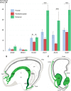
Generation of γ‐aminobutyric acid (GABA)ergic cortical interneurons in human. (A) Between 8 and 12 post‐conceptual weeks (PCW) expression of early ‘GABAergic’ genes associated with ventral telencephalic domains in rodents is also very low in the human dorsal telencephon, but ‘GABAergic’ genes expressed in late progenitor and post‐mitotic cells shower higher expression, particularly in frontal temporal cortex. (B) and (C) Interneuron migration pathways proposed to be more prominent in human than mouse, including an anterior pathway from caudal ganglionic eminence (CGE) to frontal cortex and a medial pathway from septum to frontomedial cortex.
Figure 6
Figure 6
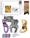
The human subplate. Summary figure showing synaptic distribution (A), neurons (B), early astroglia (C), transient circuitry (D), synapse (E) and delineation of subplate (F–I). Graphic representation of the spatial distribution of synapses (A) in the somatosensory cortex of a 15‐week‐old human fetus obtained in 23 probes each 5 μm wide, in the abscissa as a function of 100 μm (with permission from Kostović & Rakic, 1990). Golgi impregnated large neuron (B) with long smooth dendrites (with permission from Kostović et al. 2019). ‘Transient’ astroglia in the subplate (C) (with permission from Kostović et al. 2019). Electromicrograph (E) of the growth‐cone‐like dendritic profile forming a synaptic junction (arrow) in the subplate of 21‐week‐old human fetus (with permission from Kostović & Rakic, 1990). Transient circuitry of subplate (D) in a 24‐week human fetus consists of thalamus‐subplate‐thalamus circuit (red), basal forebrain (blue) and monoaminergic input (green), connections between SP neurons and transient projection to the cortical plate (with permission from Kostović & Judaš, 2007). Comparative images of subplate on Nissl (F, I), MR in vitro (G) and AChE preparations (H) show that subplate is the thickest compartment of the cerebral wall situated between cortical plate and external sagittal stratum (arrowhead) (with permission from Kostović et al. 2002).
Figure 7
Figure 7
Spatiotemporal localization of disease‐associated genes in cortical cell populations during non‐human primate brain development. (A) Weighted correlation network analysis (WGCNA) co‐expression networks (gene modules) based on 603 laser‐microdissected samples from layers of primary visual and anterior cingulate cortex from rhesus monkey brain. WGCNA was run independently at each age (columns), including six prenatal timepoints (reported in days post‐conception) spanning early to mid‐fetal development, and four postnatal timepoints from birth to young adulthood. Modules were tested for significant gene set enrichment and overlap with hypergeometric tests. Modules significantly enriched for markers of glial cell classes (P < 10−15) or cortical layers (P < 10−30) are color‐coded and annotated. The remaining modules are colored or labeled based on maximal expression in post‐mitotic (neuron‐enriched; cyan) layers or progenitor or largely non‐neuronal (WM, layer 1; orange) layers. Modules from adjacent ages with the most highly significant gene overlap (P < 10−50) are connected by gray lines. (B–D) Left: modules significantly enriched for risk genes associated with neurodevelopmental disorders (empirically corrected P < 0.1; red discs). Right: average expression pattern of genes found in at least two enriched modules. Heat maps are organized by dissected layer and age. MZ, marginal zone; CPo/CPi, outer/inner cortical plate; SP, subplate; IZ, intermediate zone; SZ, subventricular zone; VZo/VZi, outer/inner ventricular zone. (B) Genes related to primary autosomal recessive microcephaly are enriched in early non‐neuronal modules, and show maximal expression in prenatal ventricular zone. (C) Genes related to autism spectrum disorder (ASD) are enriched in modules associated with cortical neurons and show highest expression in cortical plate across development. (D) Genes related to schizophrenia show similar neuronal layer enrichment to autism genes, but restricted to postnatal ages. The figure is reproduced from Bakken et al. (2016).
Figure 8
Figure 8
Impact of neurosensory stimuli on cortical development. Neonatal fMRI reveals enhanced music processing in preterm and full‐term newborns exposed to music starting from 33 weeks post‐conceptional age until term equivalent age compared with two additional groups without music intervention; preterm infants and full‐term newborns. Each row shows in a sagittal, coronal, and axial plane, results of the PPI analysis for Original  > Tempo‐Modification conditions (P < 0.05 FWE at cluster level). (A, B) Enhanced connectivity in Preterm‐Music compared with Preterm‐Control group between right primary auditory cortex (seed) and (A) the right thalamus and (B) the left caudate nucleus and middle cingulate cortex (MCC; P < 0.01 FWE at cluster level). (C, D) Enhanced connectivity in Preterm‐Music compared with Full‐Term group between left primary auditory cortex (seed) and (C) the left superior temporal gyrus and (D) the MCC. (E) Enhanced connectivity in Preterm‐Music compared with Full‐Term group between right primary auditory cortex (seed) and (E) the left MCC cortex and left putamen. Figure adopted from Lordier et al. (2019a) NeuroImage with permission from Elsevier.

References

    1. Adam‐Darque A, Grouiller F, Vasung L, et al. (2017) fMRI‐based neuronal response to new odorants in the newborn brain. Cereb Cortex 27, 1–7. - PubMed
    1. Adorján I, Tyler T, Bhaduri A, et al. (2019) Neuroserpin expression during human brain development and in adult brain revealed by immunohistochemistry and single cell RNA sequencing. J Anat. 10.1111/joa.1293. - DOI - PMC - PubMed
    1. Alfano C, Studer M (2013) Neocortical arealization: evolution, mechanisms, and open questions. Dev Neurobiol 73, 411–447. - PubMed
    1. Al‐Jaberi N, Lindsay S, Sarma S, et al. (2015) The early fetal development of human neocortical GABAergic interneurons. Cereb Cortex 25, 631–645. - PMC - PubMed
    1. Allendoerfer K, Shatz C (1994) The subplate, a transient neocortical structure: its role in the development of connections between thalamus and cortex. Annu Rev Neurosci 17, 185–218. - PubMed