Review
. 2019 Sep 1;51(9):432-442.
doi: 10.1152/physiolgenomics.00006.2019. Epub 2019 Aug 2.

Medial prefrontal cortex in neurological diseases

Affiliations
Review

Medial prefrontal cortex in neurological diseases

Pan Xu et al. Physiol Genomics. .

Abstract

The medial prefrontal cortex (mPFC) is a crucial cortical region that integrates information from numerous cortical and subcortical areas and converges updated information to output structures. It plays essential roles in the cognitive process, regulation of emotion, motivation, and sociability. Dysfunction of the mPFC has been found in various neurological and psychiatric disorders, such as depression, anxiety disorders, schizophrenia, autism spectrum disorders, Alzheimer's disease, Parkinson's disease, and addiction. In the present review, we summarize the preclinical and clinical studies to illustrate the role of the mPFC in these neurological diseases.

Keywords: cortical region; medial prefrontal cortex; neural circuit; neurological disease; pathophysiology.


Conflict of interest statement

No conflicts of interest, financial or otherwise, are declared by the authors.

Figures

Fig. 1.
Fig. 1.
Schematic structure of different regions and layers of the medial prefrontal cortex (mPFC). The mPFC consists of four distinct neuroanatomical subregions along the dorsal to ventral axis: medial precentral area (PrCm), anterior cingulate cortex (ACC), prelimbic cortex (PL), and infralimbic cortex (IL). Its laminar organization is divided into six distinct layers (I–VI) from the superficial, and the pyramidal neurons (PN) and parvalbumin (PV) interneurons in those layers maintain an excitation/inhibition balance to achieve the normal functions of the mPFC.
Fig. 2.
Fig. 2.
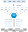
The mPFC conceived as a network. The mPFC network consists of the connections within mPFC neurons and the efferents and afferents connected with the cortical or subcortical brain regions, including BLA, Nac, MT, HTH, VTA, Hipp, and DRN et al. Under normal conditions, the network shows an adaptive response to input, which allows for normal behavioral functions such as emotion regulation, cognitive flexibility, working memory, social interaction, and motor coordination. However, in pathological conditions, altered neuron activity, neuron number, excitation-inhibition (E/I) balance, connectivity or other kinds of factors in mPFC are likely to underlie multiple types of neurological disorders, like the depression, schizophrenia, autism, Alzheimer’s, Parkinson’s, pain and addiction. Abbreviations: mPFC, medial prefrontal cortex; BLA, basolateral amygdala; Nac, nucleus accumbens; MT, midline thalamus; HTH, hypothalamus; VTA, ventral tegmental area; Hipp, hippocampus; DRN, dorsal raphe nucleus.

References

    1. Adhikari A, Topiwala MA, Gordon JA. Synchronized activity between the ventral hippocampus and the medial prefrontal cortex during anxiety. Neuron 65: 257–269, 2010. doi:10.1016/j.neuron.2009.12.002. - DOI - PMC - PubMed
    1. Andersen AH, Smith CD, Slevin JT, Kryscio RJ, Martin CA, Schmitt FA, Blonder LX. Dopaminergic modulation of medial prefrontal cortex deactivation in Parkinson depression. Parkinsons Dis 2015: 513452, 2015. doi:10.1155/2015/513452. - DOI - PMC - PubMed
    1. Arnsten AF, Wang MJ, Paspalas CD. Neuromodulation of thought: flexibilities and vulnerabilities in prefrontal cortical network synapses. Neuron 76: 223–239, 2012. doi:10.1016/j.neuron.2012.08.038. - DOI - PMC - PubMed
    1. Beesdo K, Knappe S, Pine DS. Anxiety and anxiety disorders in children and adolescents: developmental issues and implications for DSM-V. Psychiatr Clin North Am 32: 483–524, 2009. doi:10.1016/j.psc.2009.06.002. - DOI - PMC - PubMed
    1. Belujon P, Patton MH, Grace AA. Role of the prefrontal cortex in altered hippocampal-accumbens synaptic plasticity in a developmental animal model of schizophrenia. Cereb Cortex 24: 968–977, 2014. doi:10.1093/cercor/bhs380. - DOI - PMC - PubMed

Publication types

MeSH terms