. 2019 Aug 28;141(34):13442-13453.
doi: 10.1021/jacs.9b04695. Epub 2019 Aug 16.

Engineering Orthogonal Polypeptide GalNAc-Transferase and UDP-Sugar Pairs

Affiliations

Engineering Orthogonal Polypeptide GalNAc-Transferase and UDP-Sugar Pairs

Junwon Choi et al. J Am Chem Soc. .

Abstract

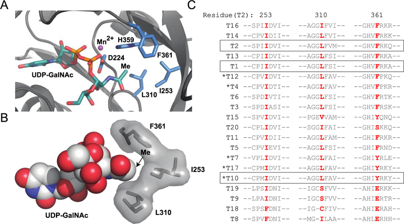
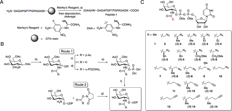
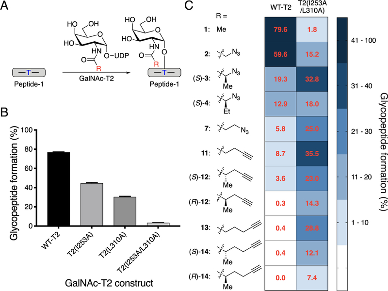
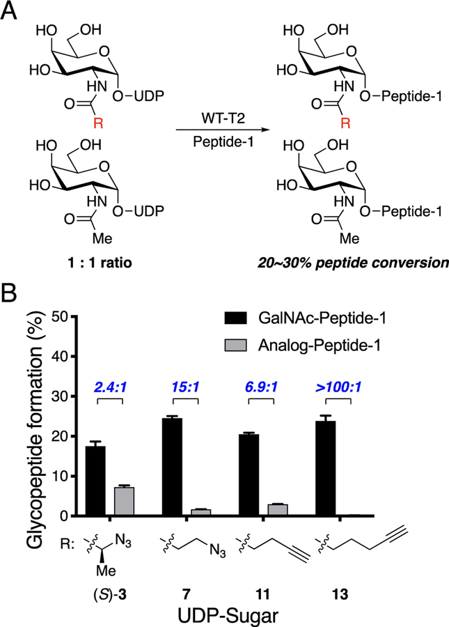
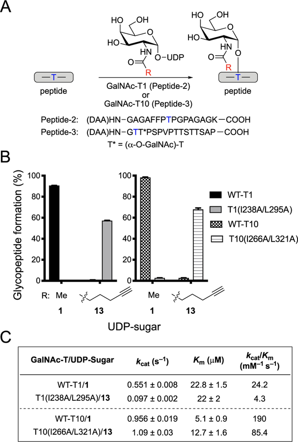
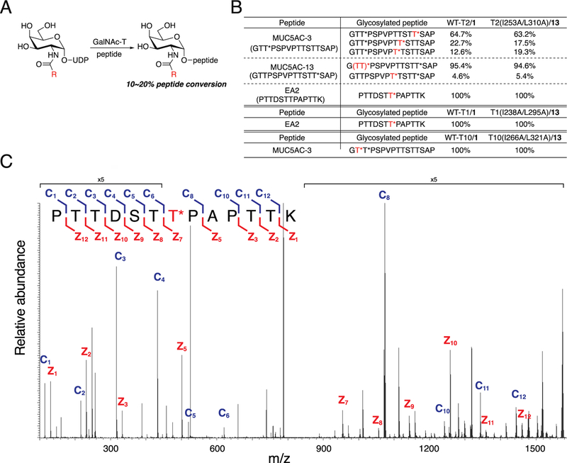
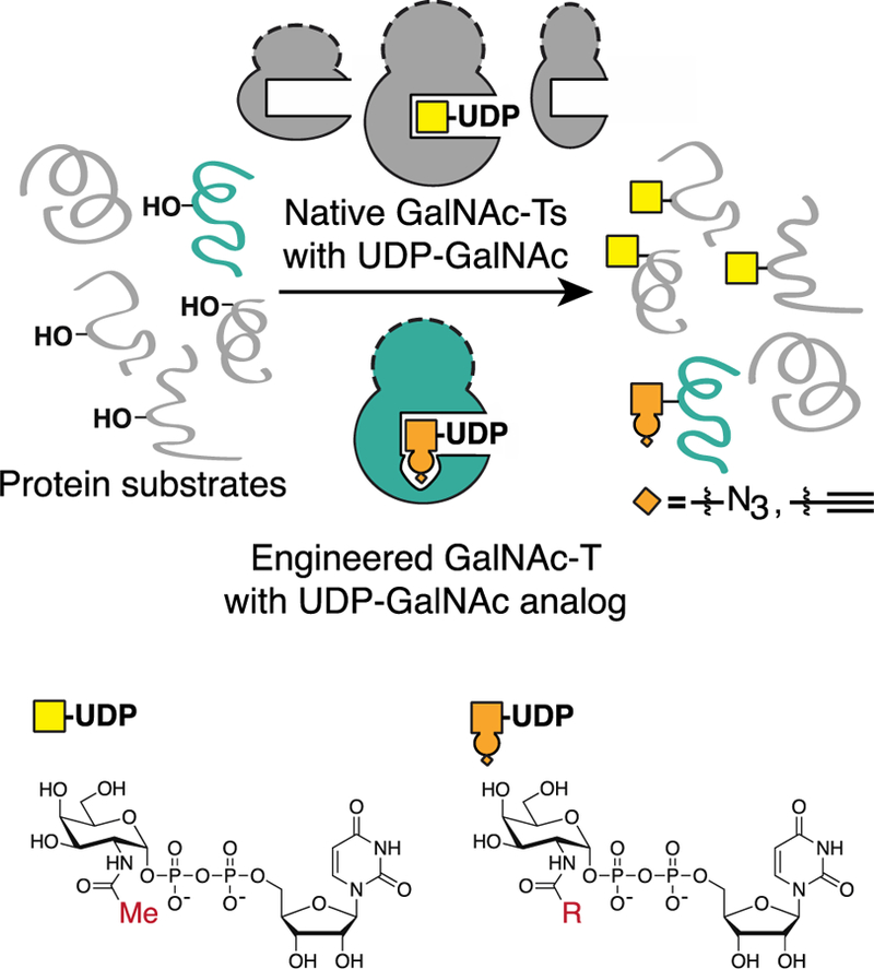
O-Linked α-N-acetylgalactosamine (O-GalNAc) glycans constitute a major part of the human glycome. They are difficult to study because of the complex interplay of 20 distinct glycosyltransferase isoenzymes that initiate this form of glycosylation, the polypeptide N-acetylgalactosaminyltransferases (GalNAc-Ts). Despite proven disease relevance, correlating the activity of individual GalNAc-Ts with biological function remains challenging due to a lack of tools to probe their substrate specificity in a complex biological environment. Here, we develop a "bump-hole" chemical reporter system for studying GalNAc-T activity in vitro. Individual GalNAc-Ts were rationally engineered to contain an enlarged active site (hole) and probed with a newly synthesized collection of 20 (bumped) uridine diphosphate N-acetylgalactosamine (UDP-GalNAc) analogs to identify enzyme-substrate pairs that retain peptide specificities but are otherwise completely orthogonal to native enzyme-substrate pairs. The approach was applicable to multiple GalNAc-T isoenzymes, including GalNAc-T1 and -T2 that prefer nonglycosylated peptide substrates and GalNAcT-10 that prefers a preglycosylated peptide substrate. A detailed investigation of enzyme kinetics and specificities revealed the robustness of the approach to faithfully report on GalNAc-T activity and paves the way for studying substrate specificities in living systems.


Conflict of interest statement

The authors declare no competing financial interest.

Figures

Figure 1.
Figure 1.
Identification of gatekeeper residues. (A) Residues within 5 Å of GalNAc methyl carbon for GalNAc-T2 (PDB ID 4D0T). Five of seven amino acids in close proximity to the GalNAc methyl contain side chains; of these, H359 and D224 coordinate Mn2+, while I253, L310, and F361 are promising hydrophobic residues. (B) Space-filling model of gatekeeper residues within 5 Å of GalNAc methyl in GalNAc-T2 (PDB ID 4D0T). (C) Amino acid sequences of human GalNAc-T1–GalNAc-T20 surrounding potential gatekeeper residues demonstrate a high degree of conservation with 13 isoenzymes containing Ile/Leu and 18 containing Ile at residues homologous to GalNAc-T2 positions 253/310 or 253, respectively. Only GalNAc-T8 and -T18 have dramatically different residues at positions corresponding to 253 and around 310 of GalNAc-T2. GalNAc-Ts used in this study are boxed. Clustal Omega was used to generate a multiple sequence alignment of the amino acid sequences corresponding to the full-length genes of human GalNAc-T1-GalNAc-T20 (Figure S2; Table S1). GalNAc-Ts are ordered based on homology, and GalNAc-Ts that predominantly prefer GalNAc-peptides are denoted with an asterisk.
Figure 2.
Figure 2.
Synthesis of a peptide substrate and a panel of UDP-GalNAc analogs. (A) Synthetic route for Peptide-1. Blue T indicates the Thr glycosylation site used by GalNAc-T2. (B) Synthetic routes for UDP-GalNAc analogs. Route 1 was used to synthesize UDP-sugars ((S)-3, (R)-3, (S)-4, (R)-4, (S)-5, (R)-5, (S)-6, (R)-6, (S)-8, (R)-8, (S)-12, (R)-12, (S)-14, (R)-14), and Route 2 was used to synthesize UDP-sugars (2, 7, 9, 10,11, 13). (C) Panel of UDP-GalNAc derivatives with azide or alkyne chemical handles. Compounds 1 and 2 are the natural substrate UDP-GalNAc and known analog UDP-GalNAz, respectively. Reagents and conditions: (a) NEt(i-Pr)2, DMF, rt; (b) R-COOH, COMU, NEt(i-Pr)2, DMF, 0 °C to rt; (c) N,N′-dimethyl-1,3-diaminopropane, THF, rt; (d) i-Pr2NPO(OAll)2, 1H-tetrazole, CH2Cl2, 0 °C, then m-CPBA, −78 °C; (e) Pd(PPh3)4, sodium p-toluenesulfinate, THF/MeOH, rt; (f) (i) uridine 5′-monophosphomorpholidate 4-morpholine-N,N′-dicyclohexylcarboxamidine salt, 1-methylimidazole hydrochloride, NEt3, DMF, rt; (ii) MeOH/water/NEt3, rt; (g) HEPES buffer (pH = 8.0), 0 °C to rt.
Figure 3.
Figure 3.
Screening GalNAc-T2 for an orthogonal enzyme−substrate pair. (A) Scheme for glycosylation reaction with Peptide-1, GalNAc-T2, and UDP-GalNAc or UDP-GalNAc analog to form glycosylated Peptide-1. Blue T indicates the Thr glycosylation site used by GalNAc-T2. (B) Glycopeptide formation by wild-type and mutant GalNAc-T2. UDP-GalNAc and Peptide-1 were incubated with GalNAc-T2 at 37 °C for 1 h, and reaction was quenched by addition of aqueous EDTA (150 mM, pH = 8.0). Percent conversion to glycopeptide product was quantified by HPLC separation and peak integration. All data represent the mean of technical triplicates, and error bars represent the standard deviation. (C) Bump−hole pair optimization for GalNAc-T2. Glycosylation by wild-type and double-mutant GalNAc-T2 was compared for UDP-GalNAc (1) and UDP-GalNAc analogs with Peptide-1. Reactions were performed and quantified as in B. Heat map (blue shading) shows percent glycosylated Peptide-1 formed by wild-type or double-mutant GalNAc-T2 with UDP-GalNAc or analogs. Red values represent the mean of technical triplicates.
Figure 4.
Figure 4.
Selectivity of wild-type GalNAc-T2 for UDP-GalNAc relative to UDP-GalNAc analogs. (A) Scheme for competition experiment between UDP-GalNAc and UDP-GalNAc analog. Wild-type GalNAc-T2 was treated with Peptide-1 and an equal ratio of UDP-GalNAc and UDP-GalNAc analog in a competition experiment, and glycosylation reactions were terminated at 20−30% glycopeptide formation. (B) Selectivity of wild-type GalNAc-T2 for UDP-GalNAc (1) over UDP-GalNAc analog ((S)-3, 7, 11, or 13) in a competition experiment. Reactions were performed as in A. UDP-sugars and Peptide-1 were incubated with GalNAc-T2 at 37 °C for 30 min, and reaction was quenched by addition of aqueous EDTA (150 mM, pH = 8.0). Percent conversion to glycopeptide product was quantified by HPLC separation and peak integration. Percent of Peptide-1 modified with GalNAc or GalNAc analog was measured, and selectivity ratio is shown in blue. All data represent the mean of technical triplicates, and error bars represent the standard deviation.
Figure 5.
Figure 5.
Orthogonal GalNAc-T and UDP-sugar pairs for GalNAc-T1 and GalNAc-T10. (A) Scheme for glycosylation reaction with GalNAc-T, peptide, and UDP-GalNAc or UDP-GalNAc analog to form glycosylated peptide. Glycosylation reactions with GalNAc-T1 utilized Peptide-2, and reactions with GalNAc-T10 utilized Peptide-3. Blue T indicates the Thr glycosylation site used by the GalNAc-T of interest. (B) Glycopeptide formation by wild-type or double-mutant GalNAc-T1 or GalNAc-T10 with UDP-GalNAc (1) or 13. Reactions were performed as in A. GalNAc-T, UDP-sugar, and peptide were incubated at 37 °C for 1 h (-T10) or 2 h (-T1), and reaction was quenched with aqueous EDTA (150 mM, pH = 8.0). All data represent the mean of technical triplicates, and error bars represent the standard deviation. (C) Kinetic parameters of wild-type and orthogonal GalNAc-T and UDP-sugar pairs. To determine Km and kcat values for UDP-GalNAc and UDP-GalNAc analogs, initial rates were measured by incubating wild-type or double-mutant GalNAc-Ts with varying concentrations of UDP-sugars and a constant concentration of acceptor peptide. For GalNAc-T1, the concentration of UDP-sugars varied from 15.6 to 500 μM, and the concentration of acceptor Peptide-2 was held at 250 μM. For GalNAc-T10, the concentration of UDP-sugars varied from 15.6 to 250 μM, and the concentration of acceptor Peptide-3 was held at 266 μM. Glycosylation was conducted at 37 °C, and three aliquots were taken within 15 min and quenched by addition of aqueous EDTA (150 mM, pH = 8.0). Products were quantified by HPLC separation and peak integration. Enzymatic kinetic parameters were obtained by nonlinear regression fitting using GraphPad Prism. All data represent the mean of technical triplicates, and error depicts the standard deviation.
Figure 6.
Figure 6.
Glycosylation of natural peptide substrates by wild-type and engineered GalNAc-T isoenzyme−substrate pairs. (A) Scheme for glycosylation reaction with GalNAc-T, natural peptide substrate, and UDP-GalNAc or UDP-GalNAc analog to form glycosylated peptide. Glycosylation reactions were terminated at 10−20% glycopeptide formation. (B) Percent of glycosylated peptide formed out of total glycosylated peptide formed. Reactions were performed as in A at 37 °C and quenched by addition of aqueous EDTA (150 mM, pH = 8.0). Naturally occurring glycopeptides MUC5AC-3 and MUC5AC-13 each contain a single GalNAc-O-Thr (T*). Red T* indicates the site of glycosylation by the GalNAc-T of interest. Glycosylation of MUC5AC-3 by GalNAc-T2 yielded a major product that was glycosylated at Thr3 by wild-type GalNAc-T2/1. The glycosite from T2(I253A/L310A)/13 could not be unambiguously assigned and was either Thr2 or Thr3, labeled (TT)*. (C) Representative MS/MS spectrum of EA2 glycosylated by T2(1253A/L310A)/13 upon fragmentation and sequencing. Fragmentation pattern of EA2 amino acid sequence to generate c ions (blue) and z ions (red) is shown.
Scheme 1.
Scheme 1.. Bump–Hole Approacha
aMutagenesis of key gatekeeper residues in the active site of a GalNAc-T introduces a “hole” in the catalytic domain of the engineered GalNAc-T that accommodates an enlarged UDP-GalNAc analog modified with a “bump” (orange circle) and chemical handle (orange diamond). The N-acyl side chain of UDP-GalNAc contains a methyl group (red Me) that is modified on the UDP-GalNAc analog to an R-group (red R), representing the bump and chemical handle. Monosaccharides are represented with colored boxes: GalNAc (yellow) and GalNAc analog (orange). The lectin domain of a GalNAc-T is represented as semicircle (dashed line).

References

    1. Vakhrushev SY; Steentoft C; Vester-Christensen MB; Bennett EP; Clausen H; Levery SB Enhanced Mass Spectrometric Mapping of the Human GalNAc-Type O-Glycoproteome with SimpleCells. Mol. Cell. Proteomics 2013, 12 (4), 932–944. - PMC - PubMed
    1. Tran DT; Zhang L; Zhang Y; Tian E; Earl LA; Ten Hagen KG Multiple Members of the UDP-GalNAc: Polypeptide N-Acetylgalactosaminyltransferase Family Are Essential for Viability in Drosophila. J. Biol. Chem 2012, 287 (8), 5243–5252. - PMC - PubMed
    1. Xu Z; Weiss A Negative Regulation of CD45 by Differential Homodimerization of the Alternatively Spliced Isoforms. Nat. Immunol 2002, 3 (8), 764–771. - PubMed
    1. Schjoldager KT-BG; Vester-Christensen MB; Bennett EP; Levery SB; Schwientek T; Yin W; Blixt O; Clausen H O-Glycosylation Modulates Proprotein Convertase Activation of Angiopoietin-like Protein 3. J. Biol. Chem 2010, 285 (47), 36293–36303. - PMC - PubMed
    1. Radhakrishnan P; Dabelsteen S; Madsen FB; Francavilla C; Kopp KL; Steentoft C; Vakhrushev SY; Olsen JV; Hansen L; Bennett EP; Woetmann A; Yin G; Chen L; Song H; Bak M; Hlady RA; Peters SL; Opavsky R; Thode C; Qvortrup K; Schjoldager KT-BG; Clausen H; Hollingsworth MA; Wandall HH Immature Truncated O-Glycophenotype of Cancer Directly Induces Oncogenic Features. Proc. Natl. Acad. Sci. U. S. A 2014, 111 (39), E4066–E4075. - PMC - PubMed

Publication types

MeSH terms