

Analysis of nickel distribution by synchrotron radiation X-ray fluorescence in nickel-induced early- and late-phase allergic contact dermatitis in Hartley guinea pigs
- PMID: 31373908
- PMCID: PMC6708687
- DOI: 10.1097/CM9.0000000000000365
Analysis of nickel distribution by synchrotron radiation X-ray fluorescence in nickel-induced early- and late-phase allergic contact dermatitis in Hartley guinea pigs
Abstract
Background: Nickel-induced allergic contact dermatitis (Ni-ACD) is a global health problem. More detailed knowledge on the skin uptake of haptens is required. This study aimed to investigate the penetration process and distribution of nickel in skin tissues with late phase and early phase of Ni-ACD to understand the mechanisms of metal allergy.
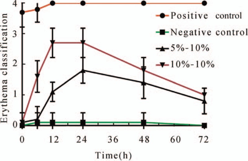
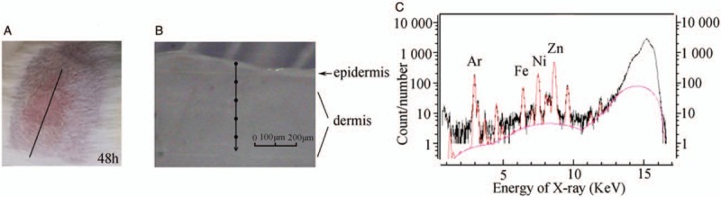
Methods: Forty Hartley guinea pigs were divided into four groups according to the NiSO4 sensitizing concentration and the NiSO4 challenged concentration: the 5% NiSO4-group, 5% to 10% (sensitization-challenge; late phase group); 10% NiSO4-group, 10% to 10% (sensitization-challenge; early-phase group); and the positive and negative controls. Pathological biopsies were performed on each group. The depth profile of nickel element concentration in the skin of guinea pigs was detected by synchrotron radiation micro X-ray fluorescence spectroscopy (SR-μ-XRF) and micro X-ray absorption near-edge spectroscopy (μ-XANES).
Results: In each section, the nickel element concentration in both the 5% NiSO4-group and 10% NiSO4-group was significantly higher than that in the negative control group. In the upper 300-μm section of skin for the early phase group, the nickel element concentration was significantly higher than that in the lower section of skin. In deeper sections (>200 μm) of skin, the concentration of nickel in the early phase group was approximately equal to that in the late phase group. The curve of the late phase group was flat, which means that the nickel element concentration was distributed uniformly by SR-μ-XRF. According to the XANES data for the 10% NiSO4 metal salt solution, structural changes occurred in the skin model sample, indicating that nickel was not present in the Ni aqueous ionic state but in the nickel-binding protein.
Conclusions: This study showed that the distribution of the nickel element concentration in ACD skin tissue was different between the early phase and late phase groups. The nickel element was not present in the Ni aqueous ionic state but bound with certain proteins to form a complex in the stratum corneum in ACD model tissue.
Figures


Similar articles
-
Zinc effects on nickel dermatitis in the guinea pig.Contact Dermatitis. 1988 Aug;19(2):98-108. doi: 10.1111/j.1600-0536.1988.tb05505.x. Contact Dermatitis. 1988. PMID: 3180790
-
Nickel deposition and penetration into the stratum corneum after short metallic nickel contact: An experimental study.Contact Dermatitis. 2019 Feb;80(2):86-93. doi: 10.1111/cod.13136. Epub 2018 Oct 29. Contact Dermatitis. 2019. PMID: 30370609
-
Experimental sensitization of guinea-pigs to nickel and patch testing with metal samples.Food Chem Toxicol. 1987 Jan;25(1):83-5. doi: 10.1016/0278-6915(87)90309-7. Food Chem Toxicol. 1987. PMID: 3817663
-
The Unique Molecular Signatures of Contact Dermatitis and Implications for Treatment.Clin Rev Allergy Immunol. 2019 Feb;56(1):1-8. doi: 10.1007/s12016-018-8685-0. Clin Rev Allergy Immunol. 2019. PMID: 29754191 Review.
-
Nickel Allergy: Epidemiology, Pathomechanism, Clinical Patterns, Treatment and Prevention Programs.Endocr Metab Immune Disord Drug Targets. 2020;20(7):992-1002. doi: 10.2174/1871530320666200128141900. Endocr Metab Immune Disord Drug Targets. 2020. PMID: 31994473 Review.
References
-
- Thyssen JP, Linneberg A, Menné T, Johansen JD. The epidemiology of contact allergy in the general population–prevalence and main findings. Contact Dermatitis 2007; 57:287–299. doi: 10.1111/j.1600-0536.2007.01220.x. - PubMed
-
- Thyssen JP, Linneberg A, Menné T, Johansen JD. Quantitative aspects of nickel dermatitis. Sensitization and eliciting threshold concentrations. Sci Total Environ 1994; 148:275–281. doi: 10.1016/0048-9697(94)90403-0. - PubMed
-
- Spiewak R, Pietowska J, Curzytek K. Nickel: a unique allergen–from molecular structure to European legislation. Expert Rev Clin Immunol 2007; 3:851–859. doi: 10.1586/1744666X.3.6.851. - PubMed
-
- Ahlström MG, Menné T, Thyssen JP, Johansen JD. Nickel allergy in a Danish population 25 years after the first nickel regulation. Contact Dermatitis 2017; 76:325–332. doi: 10.1111/cod.12782. - PubMed
-
- Staton I, Ma R, Evans N, Hutchinson RW, McLeod CW, Gawkrodger DJ. Dermal nickel exposure associated with coin handling and in various occupational settings: assessment using a newly developed finger immersion method. Br J Dermatol 2006; 154:658–664. doi: 10.1111/j.1365-2133.2006.07128.x. - PubMed
MeSH terms
Substances
LinkOut - more resources
Full Text Sources
Research Materials
