. 2019 Aug 2;14(8):e0220573.
doi: 10.1371/journal.pone.0220573. eCollection 2019.

The right ventricular fibroblast secretome drives cardiomyocyte dedifferentiation

Affiliations

The right ventricular fibroblast secretome drives cardiomyocyte dedifferentiation

Danielle R Bruns et al. PLoS One. .

Abstract

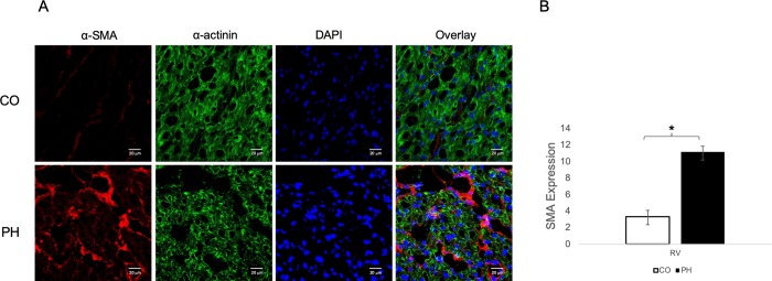
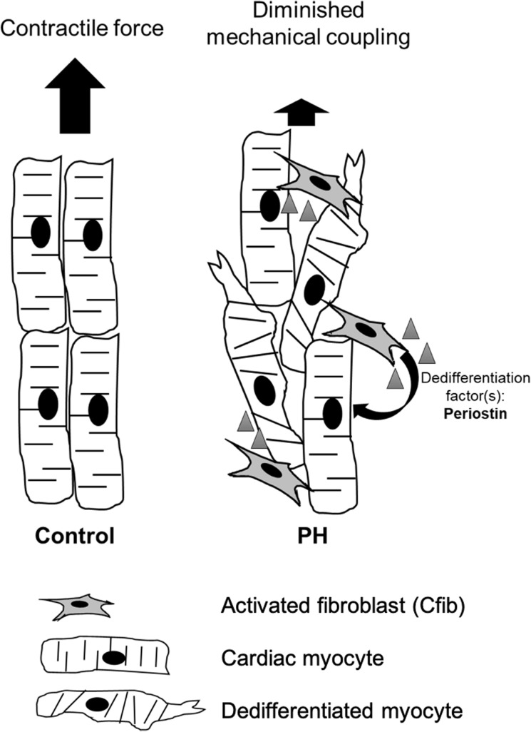
Rationale: In virtually all models of heart failure, prognosis is determined by right ventricular (RV) function; thus, understanding the cellular mechanisms contributing to RV dysfunction is critical. Whole organ remodeling is associated with cell-specific changes, including cardiomyocyte dedifferentiation and activation of cardiac fibroblasts (Cfib) which in turn is linked to disorganization of cytoskeletal proteins and loss of sarcomeric structures. However, how these cellular changes contribute to RV function remains unknown. We've previously shown significant organ-level RV dysfunction in a large animal model of pulmonary hypertension (PH) which was not mirrored by reduced function of isolated cardiomyocytes. We hypothesized that factors produced by the endogenous Cfib contribute to global RV dysfunction by generating a heterogeneous cellular environment populated by dedifferentiated cells.

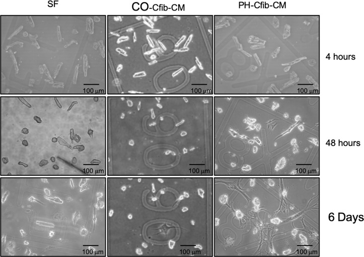
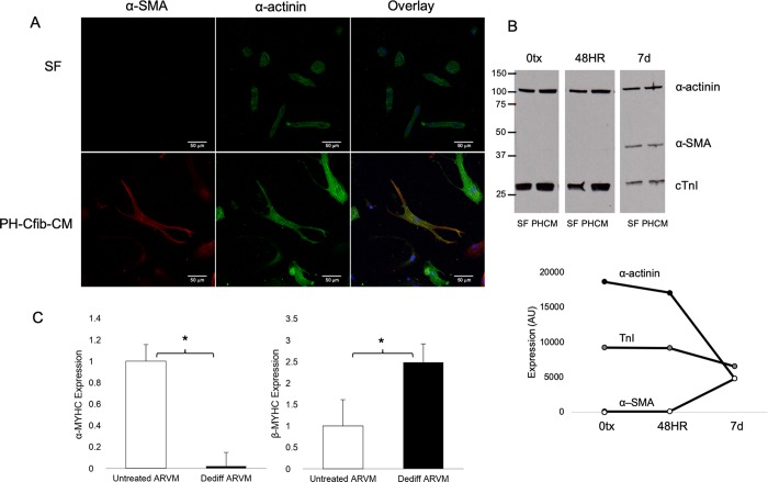
Objective: To determine the effect of Cfib conditioned media (CM) from the PH calf (PH-CM) on adult rat ventricular myocytes (ARVM) in culture.

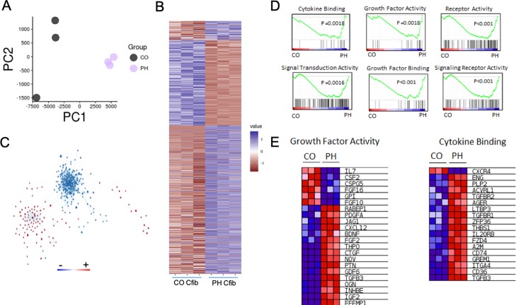
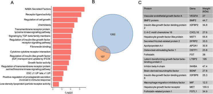
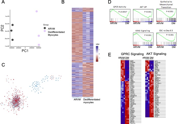
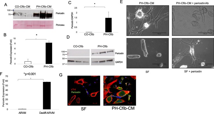
Methods and results: Brief exposure (<2 days) to PH-CM results in rapid, marked dedifferentiation of ARVM to a neonatal-like phenotype exhibiting spontaneous contractile behavior. Dedifferentiated cells maintain viability for over 30 days with continued expression of cardiomyocyte proteins including TnI and α-actinin yet exhibit myofibroblast characteristics including expression of α-smooth muscle actin. Using a bioinformatics approach to identify factor(s) that contribute to dedifferentiation, we found activation of the PH Cfib results in a unique transcriptome correlating with factors both in the secretome and with activated pathways in the dedifferentiated myocyte. Further, we identified upregulation of periostin in the Cfib and CM, and demonstrate that periostin is sufficient to drive cardiomyocyte dedifferentiation.

Conclusions: These data suggest that paracrine factor(s) released by Cfib from the PH calf signal a phenotypic transformation in a population of cardiomyocytes that likely contributes to RV dysfunction. Therapies targeting this process, such as inhibition of periostin, have the potential to prevent RV dysfunction.


Conflict of interest statement

The authors have declared that no competing interests exist.

Figures

Fig 1
Fig 1. Pulmonary hypertension (PH) induces myocyte disarray in vivo.
A) Representative images of mid-RV sections stained for α-SMA (red) and α-actinin (green). RV from control (CO) calves express little α-SMA and are highly organized. However, RV from PH-exposed (PH) calves express higher α-SMA and the normal organized structure of cardiac tissue is disrupted in areas expressing high α-SMA. DAPI (blue) identifies nuclei. Images were taken at 40X, with Zoom at 63X on a Zeiss Laser Scanning Microscope 780. B) Quantification of α-SMA in RV of control and PH-exposed calves (n = 12 CO; n = 12 PH).
Fig 2
Fig 2. Cardiac fibroblast conditioned media (CM) from calves with pulmonary hypertension (PH) induced right ventricular dysfunction stimulates myocyte dedifferentiation.
Adult rat ventricular myocytes (ARVM) were plated on etched coverslips to allow visual tracking of the same cell over time and incubated with serum-free (SF) media or CM derived from cardiac fibroblasts from calves exposed to control (CO) or PH conditions. By 48 hours myocytes in PH-Cfib-CM show signs of flattening, loss of striations, and branching of cellular processes. CM generated from control calf fibroblasts did not induce significant dedifferentiation.
Fig 3
Fig 3. Dedifferentiated adult myocytes retain expression of cardiac markers.
A) Immunofluorescent staining of dedifferentiated myocytes after 7 days SF or PH-CM exposure. Dedifferentiated cells still express α-actinin (green), while also starting to express α-SMA (red). Cells in SF media are less viable by 7 days, and do not dedifferentiate. Images were taken on a Zeiss Laser Scanning Microscope 780 at 40x objective. B) Protein expression of sarcomeric α-actinin, cardiac troponin I (cTnI) and α-SMA in myocytes exposed to SF media or PH-CM for 48 hours or 7 days. CD) Dedifferentiated myocytes activate the fetal gene program. By 7 days in culture, cells exposed to PH-CM demonstrate downregulated SERCA and α-MYHC expression. Expression of the fetal gene program was assessed by qRT-PCR using validated primers. Data are expressed as ΔCt to allow comparison across all groups. Higher reflects lower expression. n = 2 at each time-point. *p<0.05 compared to 0tx.
Fig 4
Fig 4. RNAseq analysis of control (CO) and pulmonary hypertension (PH) cardiac fibroblasts.
A) PCA analysis of RNAseq data from CO and PH fibroblasts. B) Heatmap of gene expression comparing control fibroblasts to pulmonary hypertension fibroblasts. Genes with a significant difference of 0.05> were selected. Expression values were scaled from -1 to 1, similarity was calculated using an euclidean distance and clustered using a centroid linkage k-means method. C) Network of transcriptional changes comparing CO and PH fibroblasts. We displayed differentially enriched pathways from GSEA as enrichment networks, in which each node is a pathway, and each link between nodes is a common gene. The size of the nodes indicates the number of genes found in our data that are in the pathway. Red coloring indicates pathways enriched in the PH group, whereas blue indicates pathways enriched in the CO group. The CO and PH networks are independent, suggesting significant divergence of in the signaling cascades between these two groups of cells. D) Select enrichment plots highlighting the enriched pathways identified by GSEA. E) Two representative heatmaps for the Growth Factor Activity and Cytokine Binding pathways showing the genes responsible for the enrichment.
Fig 5
Fig 5. Conditioned media pathway analysis of the extracellular signaling molecules.
A) Pathway analysis of signaling molecules upregulated in the PH-CM. B) Comparison of the upregulated genes in the PH Cfib transcriptome (blue circle) with significantly enhanced proteins in the PH-CM (orange circle). Of the 137 secreted proteins found enriched in the conditioned media, the transcripts of 34 of these were also found increased in the transcriptome of the PH fibroblasts. C) Representative list of 16 of the 34 proteins identified as enriched in the PH conditioned media with known signaling properties that were also found upregulated in the PH Cfib transcriptome. The human orthologues are displayed in the table and were then used for pathway analysis.
Fig 6
Fig 6. Dedifferentiated adult ventricular myocyte (ARVM) RNAseq.
A) PCA plot of ARVMs treated with and without conditioned media from PH fibroblasts. B) Heatmap of gene expression comparing control, freshly isolated ARVMs to ARVMs treated with PH Fibroblast conditioned media. Genes with a significant difference of <0.05 were selected. Expression values were scaled from -1 to 1, similarity was calculated using an euclidean distance and clustered using a centroid linkage k-means method. C) Network of transcriptional changes. We displayed differentially enriched pathways from GSEA as enrichment networks in which each node is a pathway, and each link between nodes is a common gene. The size of the nodes indicates the number of genes found in our data that are in the pathway. Red coloring indicates pathways enriched in the ARVM PH group, whereas blue indicates pathways enriched in the control ARVM group. D) Select enrichment plots highlighting the enriched pathways identified by GSEA. E) Representative heat maps for G-protein coupled receptor, downstream signaling pathways (GPCR signaling) and AKT signaling pathways showing the genes responsible for enrichment.
Fig 7
Fig 7. Periostin drives adult myocyte dedifferentiation.
A) Periostin is significantly expressed in the PH-CM compared to CO-CM. B) Periostin RNA and C,D) protein expression are significantly higher in PH Cfib compared to CO. E) Immunodepletion of periostin from the PH-CM attenuates dedifferentiation, while addition of recombinant periostin to SF media stimulates dedifferentiation. F) Periostin expression is robustly upregulated in the dedifferentiated myocytes compared to freshly isolated ARVM. G) Periostin protein expression is upregulated in dedifferentiated cells compared to SF-treated. Periostin RNA and protein expression were assessed by qRT-PCR and immunoblotting, respectively. Experiments were performed in 3 PH and 2 CO calves in biological triplicate. *p<0.05 by student’s t-test.
Fig 8
Fig 8. Working model of Cfib-mediated disruption of sarcomeric organization and loss of contractile function.
Identification of additional secreted factor(s) which drive dedifferentiation and myocyte plasticity will improve therapeutic strategies.

References

    1. Hegewisch S, Weh HJ, Hossfeld DK. TNF-induced cardiomyopathy. Lancet. 1990;335(8684):294–5. - PubMed
    1. Beohar N, Rapp J, Pandya S, Losordo DW. Rebuilding the damaged heart: the potential of cytokines and growth factors in the treatment of ischemic heart disease. J Am Coll Cardiol. 2010;56(16):1287–97. 10.1016/j.jacc.2010.05.039 - DOI - PMC - PubMed
    1. Gonzalez A, Ravassa S, Beaumont J, Lopez B, Diez J. New targets to treat the structural remodeling of the myocardium. J Am Coll Cardiol. 2011;58(18):1833–43. 10.1016/j.jacc.2011.06.058 - DOI - PubMed
    1. Liehn EA, Postea O, Curaj A, Marx N. Repair after myocardial infarction, between fantasy and reality: the role of chemokines. J Am Coll Cardiol. 2011;58(23):2357–62. 10.1016/j.jacc.2011.08.034 - DOI - PubMed
    1. Batlle M, Perez-Villa F, Lazaro A, Garcia-Pras E, Vallejos I, Sionis A, et al. Decreased expression of thrombospondin-1 in failing hearts may favor ventricular remodeling. Transplant Proc. 2009;41(6):2231–3. 10.1016/j.transproceed.2009.06.009 - DOI - PubMed

Publication types

MeSH terms