Review
. 2019 Aug 1;105(2):237-257.
doi: 10.1016/j.ajhg.2019.06.005.

DNA Damage and Associated DNA Repair Defects in Disease and Premature Aging

Affiliations
Review

DNA Damage and Associated DNA Repair Defects in Disease and Premature Aging

Vinod Tiwari et al. Am J Hum Genet. .

Abstract

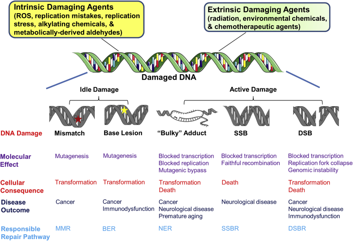
Genetic information is constantly being attacked by intrinsic and extrinsic damaging agents, such as reactive oxygen species, atmospheric radiation, environmental chemicals, and chemotherapeutics. If DNA modifications persist, they can adversely affect the polymerization of DNA or RNA, leading to replication fork collapse or transcription arrest, or can serve as mutagenic templates during nucleic acid synthesis reactions. To combat the deleterious consequences of DNA damage, organisms have developed complex repair networks that remove chemical modifications or aberrant base arrangements and restore the genome to its original state. Not surprisingly, inherited or sporadic defects in DNA repair mechanisms can give rise to cellular outcomes that underlie disease and aging, such as transformation, apoptosis, and senescence. In the review here, we discuss several genetic disorders linked to DNA repair defects, attempting to draw correlations between the nature of the accumulating DNA damage and the pathological endpoints, namely cancer, neurological disease, and premature aging.


Figures

Figure 1
Figure 1
MMR and BER (A) Replication infidelity leads to nucleotide mismatches (red star) or indels (not shown). MutS homologs (MSH) and MutL homologs (MLH) interact to carry out mismatch recognition. Pre-existing nicks in the newly synthesized strand or generated either 5′ (depicted) or 3′ (not shown) facilitate recruitment of an excision nuclease, such as EXO1. Following mismatch excision, the gap is filled, and the remaining nick sealed to complete the MMR response. See text for further details. (B) Classic BER is initiated by base damage (yellow star) removal, creating an AP site (yellow circle). AP sites can be processed by multiple enzymes but are the main substrate of APE1. Following incision at the AP damage, the break termini are processed and the gap filled. Depending on the nature of the substrate and the cellular environment, repair synthesis involves either short-patch or long-patch events. The final nick is sealed to complete the response. See text for further details. Figures were created using artwork of Servier Medical Art and Chemdraw.
Figure 2
Figure 2
NER NER is divided into two sub-pathways: GG-NER (left) and TC-NER (right). In GG-NER, DNA helix-distorting lesions are recognized by the collaborative effort of XPC, RAD23B, and CENT2, sometimes with the assistance of UV-DDB. In TC-NER, blocking lesions are revealed by arrest of an RNA polymerase at the site of damage. Persistent RNA polymerase stalling leads to recruitment of the TC-repair factors, namely CSB and CSA. Following recognition, lesion verification, damage excision, repair synthesis, and nick ligation are shared biochemical processes. See text for further details.
Figure 3
Figure 3
SSBR and DSBR (A) DNA SSBs can be generated through the action of ROS (direct) or by intended (indirect) or failed (TOP1) enzymatic processing events. PARP1 plays an important role in the resolution of direct SSBs, but may also participate in the response to other forms of DNA SSBs. Following recognition by the appropriate protein(s), specific processing enzymes are called upon to resolve abnormal 5′ or 3′ ends to permit repair synthesis and nick ligation. See text for further details. (B) DSBs can arise via several mechanisms, with two-ended DSBs being depicted. Depending on cell cycle and other factors not fully understood, DSB recognition is carried out by the Ku complex (left), PARP1 (center), or HR factors, such as ATM and the MRN complex. End processing prepares the DSB for realignment, potential gap filling, and ligation in either NHEJ event (classic or alternative), or for strand exchange, the formation of a D-loop, recombination, and eventual resolution in the case of HR. See text for further details.
Figure 4
Figure 4
Sources, Consequences, and Clinical Outcomes of DNA Damage DNA damage can arise from intrinsic or extrinsic agents or events, some of which are highlighted. DNA damaging agents can generate a variety of lesions, such as mismatched nucleotides, base lesions, bulky (helix-distorting) adducts, SSBs, or DSBs. These damages, depending on their chemical make-up or effect on transcription/replication can cause specific molecular effects, such as mutagenesis, blocked polymerization events, or genomic instability. The molecular outcomes can have specific cellular consequences, including transformation or cell death. Certain cellular events underlie disease outcomes, such as cancer, neurological disease, or premature aging. To prevent these deleterious end-points, cells have evolved a set of DNA repair mechanisms, shown at the bottom. See text for details.

References

    1. Lindahl T. Instability and decay of the primary structure of DNA. Nature. 1993;362:709–715. - PubMed
    1. Loeb L.A., Preston B.D. Mutagenesis by apurinic/apyrimidinic sites. Annu. Rev. Genet. 1986;20:201–230. - PubMed
    1. Wang W., Walmacq C., Chong J., Kashlev M., Wang D. Structural basis of transcriptional stalling and bypass of abasic DNA lesion by RNA polymerase II. Proc. Natl. Acad. Sci. USA. 2018;115:E2538–E2545. - PMC - PubMed
    1. Catalano M.J., Liu S., Andersen N., Yang Z., Johnson K.M., Price N.E., Wang Y., Gates K.S. Chemical structure and properties of interstrand cross-links formed by reaction of guanine residues with abasic sites in duplex DNA. J. Am. Chem. Soc. 2015;137:3933–3945. - PMC - PubMed
    1. Rogozin I.B., Pavlov Y.I., Goncearenco A., De S., Lada A.G., Poliakov E., Panchenko A.R., Cooper D.N. Mutational signatures and mutable motifs in cancer genomes. Brief. Bioinform. 2018;19:1085–1101. - PMC - PubMed

Publication types