Regulation of the chemotaxis histidine kinase CheA: A structural perspective
- PMID: 31374212
- PMCID: PMC7212787
- DOI: 10.1016/j.bbamem.2019.183030
Regulation of the chemotaxis histidine kinase CheA: A structural perspective
Abstract
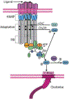
Bacteria sense and respond to their environment through a highly conserved assembly of transmembrane chemoreceptors (MCPs), the histidine kinase CheA, and the coupling protein CheW, hereafter termed "the chemosensory array". In recent years, great strides have been made in understanding the architecture of the chemosensory array and how this assembly engenders sensitive and cooperative responses. Nonetheless, a central outstanding question surrounds how receptors modulate the activity of the CheA kinase, the enzymatic output of the sensory system. With a focus on recent advances, we summarize the current understanding of array structure and function to comment on the molecular mechanism by which CheA, receptors and CheW generate the high sensitivity, gain and dynamic range emblematic of bacterial chemotaxis. The complexity of the chemosensory arrays has motivated investigation with many different approaches. In particular, structural methods, genetics, cellular activity assays, nanodisc technology and cryo-electron tomography have provided advances that bridge length scales and connect molecular mechanism to cellular function. Given the high degree of component integration in the chemosensory arrays, we ultimately aim to understand how such networked molecular interactions generate a whole that is truly greater than the sum of its parts. This article is part of a Special Issue entitled: Molecular biophysics of membranes and membrane proteins.
Keywords: Enzymology; Histidine kinase; Membrane proteins; Phosphorelay; Protein structure; Signal transduction.
Copyright © 2019. Published by Elsevier B.V.
Figures


Similar articles
-
Regulatory Role of an Interdomain Linker in the Bacterial Chemotaxis Histidine Kinase CheA.J Bacteriol. 2018 Apr 24;200(10):e00052-18. doi: 10.1128/JB.00052-18. Print 2018 May 15. J Bacteriol. 2018. PMID: 29483161 Free PMC article.
-
Noncritical Signaling Role of a Kinase-Receptor Interaction Surface in the Escherichia coli Chemosensory Core Complex.J Mol Biol. 2018 Mar 30;430(7):1051-1064. doi: 10.1016/j.jmb.2018.02.004. Epub 2018 Feb 14. J Mol Biol. 2018. PMID: 29453948 Free PMC article.
-
Identification of a Kinase-Active CheA Conformation in Escherichia coli Chemoreceptor Signaling Complexes.J Bacteriol. 2019 Nov 5;201(23):e00543-19. doi: 10.1128/JB.00543-19. Print 2019 Dec 1. J Bacteriol. 2019. PMID: 31501279 Free PMC article.
-
Bacterial chemotaxis coupling protein: Structure, function and diversity.Microbiol Res. 2019 Feb;219:40-48. doi: 10.1016/j.micres.2018.11.001. Epub 2018 Nov 6. Microbiol Res. 2019. PMID: 30642465 Review.
-
Studying bacterial chemosensory array with CryoEM.Biochem Soc Trans. 2021 Nov 1;49(5):2081-2089. doi: 10.1042/BST20210080. Biochem Soc Trans. 2021. PMID: 34495335 Free PMC article. Review.
Cited by
-
How advances in cryo-electron tomography have contributed to our current view of bacterial cell biology.J Struct Biol X. 2022 Feb 26;6:100065. doi: 10.1016/j.yjsbx.2022.100065. eCollection 2022. J Struct Biol X. 2022. PMID: 35252838 Free PMC article.
-
Chemotaxis and Shorter O-Antigen Chain Length Contribute to the Strong Desiccation Tolerance of a Food-Isolated Cronobacter sakazakii Strain.Front Microbiol. 2022 Jan 4;12:779538. doi: 10.3389/fmicb.2021.779538. eCollection 2021. Front Microbiol. 2022. PMID: 35058898 Free PMC article.
-
Coordinated regulation of chemotaxis and resistance to copper by CsoR in Pseudomonas putida.Elife. 2025 Apr 8;13:RP100914. doi: 10.7554/eLife.100914. Elife. 2025. PMID: 40197389 Free PMC article.
-
Xanthomonas oryzae Pv. oryzicola Response Regulator VemR Is Co-opted by the Sensor Kinase CheA for Phosphorylation of Multiple Pathogenicity-Related Targets.Front Microbiol. 2022 Jun 9;13:928551. doi: 10.3389/fmicb.2022.928551. eCollection 2022. Front Microbiol. 2022. PMID: 35756024 Free PMC article.
-
Allostery and protein plasticity: the keystones for bacterial signaling and regulation.Biophys Rev. 2021 Nov 10;13(6):943-953. doi: 10.1007/s12551-021-00892-9. eCollection 2021 Dec. Biophys Rev. 2021. PMID: 35059019 Free PMC article. Review.
References
-
- Armitage JP, Bacterial tactic responses, Adv Microb Physiol, 41 (1999) 229–289. - PubMed
Publication types
MeSH terms
Substances
Grants and funding
LinkOut - more resources
Full Text Sources

