

Cardiac Cx43 and ECM Responses to Altered Thyroid Status Are Blunted in Spontaneously Hypertensive versus Normotensive Rats
- PMID: 31374823
- PMCID: PMC6696036
- DOI: 10.3390/ijms20153758
Cardiac Cx43 and ECM Responses to Altered Thyroid Status Are Blunted in Spontaneously Hypertensive versus Normotensive Rats
Abstract
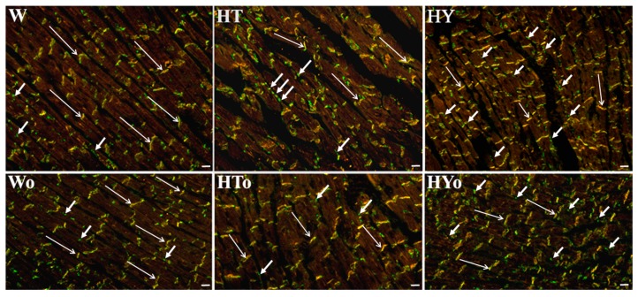
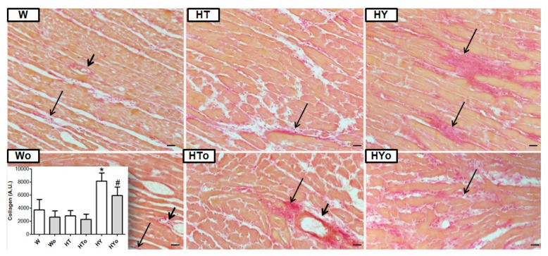
Heart function and its susceptibility to arrhythmias are modulated by thyroid hormones (THs) but the responsiveness of hypertensive individuals to thyroid dysfunction is elusive. We aimed to explore the effect of altered thyroid status on crucial factors affecting synchronized heart function, i.e., connexin-43 (Cx43) and extracellular matrix proteins (ECM), in spontaneously hypertensive rats (SHRs) compared to normotensive Wistar Kyoto rats (WKRs). Basal levels of circulating THs were similar in both strains. Hyperthyroid state (HT) was induced by injection of T3 (0.15 mg/kg b.w. for eight weeks) and hypothyroid state (HY) by the administration of methimazol (0.05% for eight weeks). The possible benefit of omega-3 polyunsaturated fatty acids (Omacor, 200 mg/kg for eight weeks) intake was examined as well. Reduced levels of Cx43 in SHRs were unaffected by alterations in THs, unlike WKRs, in which levels of Cx43 and its phosphorylated form at serine368 were decreased in the HT state and increased in the HY state. This specific Cx43 phosphorylation, attributed to enhanced protein kinase C-epsilon signaling, was also increased in HY SHRs. Altered thyroid status did not show significant differences in markers of ECM or collagen deposition in SHRs. WKRs exhibited a decrease in levels of profibrotic transforming growth factor β1 and SMAD2/3 in HT and an increase in HY, along with enhanced interstitial collagen. Short-term intake of omega-3 polyunsaturated fatty acids did not affect any targeted proteins significantly. Key findings suggest that myocardial Cx43 and ECM responses to altered thyroid status are blunted in SHRs compared to WKRs. However, enhanced phosphorylation of Cx43 at serine368 in hypothyroid SHRs might be associated with preservation of intercellular coupling and alleviation of the propensity of the heart to malignant arrhythmias.
Keywords: connexin-43; extracellular matrix; heart; spontaneously hypertensive rats; thyroid hormones.
Conflict of interest statement
The authors declare no conflict of interest.
Figures


Similar articles
-
Cardiac connexin-43 and PKC signaling in rats with altered thyroid status without and with omega-3 fatty acids intake.Physiol Res. 2016 Sep 19;65 Suppl 1:S77-90. doi: 10.33549/physiolres.933413. Physiol Res. 2016. PMID: 27643942
-
Distinct Cardiac Connexin-43 Expression in Hypertrophied and Atrophied Myocardium May Impact the Vulnerability of the Heart to Malignant Arrhythmias. A Pilot Study.Physiol Res. 2023 Jun 9;72(S1):S37-S45. doi: 10.33549/physiolres.935025. Physiol Res. 2023. PMID: 37294117 Free PMC article.
-
Altered thyroid status affects myocardial expression of connexin-43 and susceptibility of rat heart to malignant arrhythmias that can be partially normalized by red palm oil intake.Histochem Cell Biol. 2017 Jan;147(1):63-73. doi: 10.1007/s00418-016-1488-6. Epub 2016 Sep 6. Histochem Cell Biol. 2017. PMID: 27600718
-
Effects of long-term thyroid hormone level alterations, n-3 polyunsaturated fatty acid supplementation and statin administration in rats.Physiol Res. 2014;63(Suppl 1):S119-31. doi: 10.33549/physiolres.932623. Physiol Res. 2014. PMID: 24564652 Review.
-
Protection of cardiac cell-to-cell coupling attenuate myocardial remodeling and proarrhythmia induced by hypertension.Physiol Res. 2016 Sep 19;65 Suppl 1:S29-42. doi: 10.33549/physiolres.933391. Physiol Res. 2016. PMID: 27643938 Review.
Cited by
-
Does Myocardial Atrophy Represent Anti-Arrhythmic Phenotype?Biomedicines. 2022 Nov 4;10(11):2819. doi: 10.3390/biomedicines10112819. Biomedicines. 2022. PMID: 36359339 Free PMC article. Review.
-
New aspects of endocrine control of atrial fibrillation and possibilities for clinical translation.Cardiovasc Res. 2021 Jun 16;117(7):1645-1661. doi: 10.1093/cvr/cvab080. Cardiovasc Res. 2021. PMID: 33723575 Free PMC article. Review.
-
Cardiac Cx43 Signaling Is Enhanced and TGF-β1/SMAD2/3 Suppressed in Response to Cold Acclimation and Modulated by Thyroid Status in Hairless SHRM.Biomedicines. 2022 Jul 14;10(7):1707. doi: 10.3390/biomedicines10071707. Biomedicines. 2022. PMID: 35885012 Free PMC article.
-
The treatment with trandolapril and losartan attenuates pressure and volume overload alternations of cardiac connexin-43 and extracellular matrix in Ren-2 transgenic rats.Sci Rep. 2023 Nov 27;13(1):20923. doi: 10.1038/s41598-023-48259-2. Sci Rep. 2023. PMID: 38017033 Free PMC article.
-
Myocardial Adaptation in Pseudohypoxia: Signaling and Regulation of mPTP via Mitochondrial Connexin 43 and Cardiolipin.Cells. 2019 Nov 17;8(11):1449. doi: 10.3390/cells8111449. Cells. 2019. PMID: 31744200 Free PMC article. Review.
References
MeSH terms
Substances
Grants and funding
LinkOut - more resources
Full Text Sources
Medical
Molecular Biology Databases
