

Anti-Tumor Effects of Vitamin B2, B6 and B9 in Promonocytic Lymphoma Cells
- PMID: 31374832
- PMCID: PMC6696026
- DOI: 10.3390/ijms20153763
Anti-Tumor Effects of Vitamin B2, B6 and B9 in Promonocytic Lymphoma Cells
Abstract
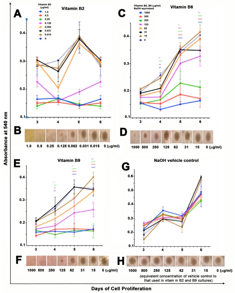
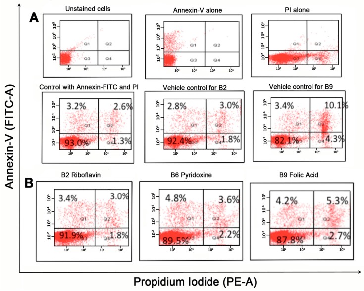
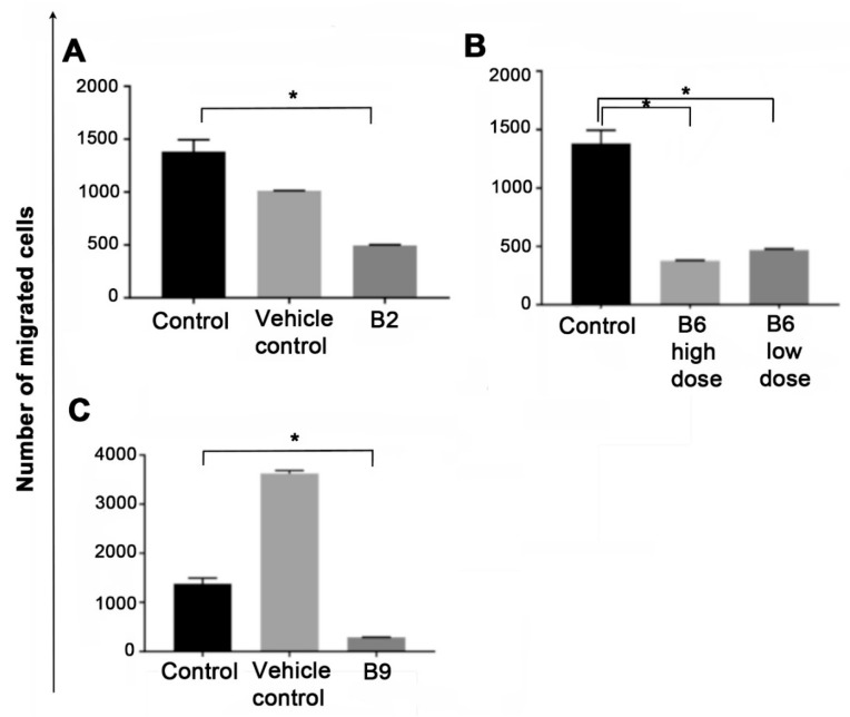
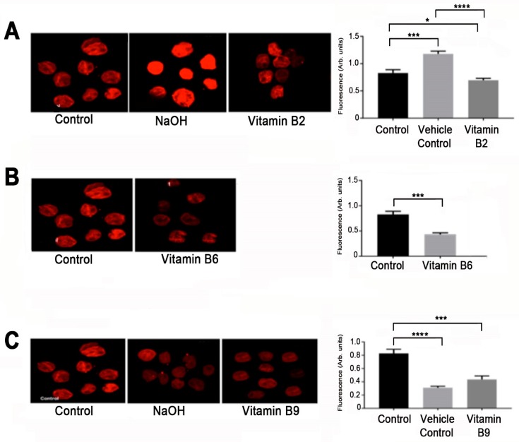
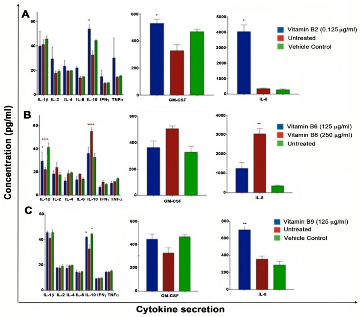
Chronic inflammation can lead to tumour initiation and progression. Vitamin B complex has the ability to regulate the immune response and, therefore, inflammation but many of the mechanistic and molecular processes involved in this regulation are still not fully understood. This study sought to determine some of these processes by studying the effects of vitamin B2 (riboflavin) B6 (pyridoxine) and B9 (folic acid) on un-differentiated pro-monocytic lymphoma cells in regard to their ability to alter the proliferation, migration, apoptosis, cytokines and expression levels of programmed death ligand 1. We show that vitamin B2, B6 and B9, on pro-monocytic lymphoma cells exerted an anti-tumorigenic effect. This data could form the basis for future studies in using vitamin B supplementation to reduce cancer cell growth in vivo.
Keywords: U937 cell line; folate; pro-monocytes; pyridoxine; riboflavin; vitamin B complex; vitamin B2; vitamin B6; vitamin B9.
Conflict of interest statement
The authors declare no conflict of interest.
Figures
Similar articles
-
Influence of folic acid, vitamin B2 and B6 supplementation on feed intake, body and organs weight, and liver fatty acids composition in rats subjected to severe protein deprivation.Pol J Vet Sci. 2006;9(3):185-90. Pol J Vet Sci. 2006. PMID: 17020013
-
Effect of restricted feed intake and addition of vitamins B2 and B6 and folic Acid on the liver concentration of zinc and copper in rats.Biol Trace Elem Res. 2004 Apr;98(1):85-94. doi: 10.1385/BTER:98:1:85. Biol Trace Elem Res. 2004. PMID: 15051903
-
[Influence of pyridoxin and riboflavin on free methionine in liver in guinea pigs].Arch Maragliano Patol Clin. 1955 Jun;10(3):855-60. Arch Maragliano Patol Clin. 1955. PMID: 13269230 Italian. No abstract available.
-
Folate and vitamin B12 metabolism: overview and interaction with riboflavin, vitamin B6, and polymorphisms.Food Nutr Bull. 2008 Jun;29(2 Suppl):S5-16; discussion S17-9. doi: 10.1177/15648265080292S103. Food Nutr Bull. 2008. PMID: 18709878 Review.
-
Nutritional effects of oral contraceptive use: a review.J Reprod Med. 1980 Oct;25(4):150-6. J Reprod Med. 1980. PMID: 7001015 Review.
Cited by
-
Study on the correlation between B vitamins and breast cancer.Cancer Cell Int. 2023 Feb 9;23(1):22. doi: 10.1186/s12935-023-02860-7. Cancer Cell Int. 2023. PMID: 36759846 Free PMC article.
-
Vitamin B6 in Health and Disease.Nutrients. 2021 Sep 17;13(9):3229. doi: 10.3390/nu13093229. Nutrients. 2021. PMID: 34579110 Free PMC article. Review.
-
Genetically predicted vitamins supplementation and risk of skin cancers: a Mendelian randomization study.Discov Oncol. 2025 Feb 19;16(1):204. doi: 10.1007/s12672-025-01905-9. Discov Oncol. 2025. PMID: 39969704 Free PMC article.
-
Molecular mechanisms and therapeutic effects of different vitamins and minerals in COVID-19 patients.J Trace Elem Med Biol. 2022 Sep;73:127044. doi: 10.1016/j.jtemb.2022.127044. Epub 2022 Jul 20. J Trace Elem Med Biol. 2022. PMID: 35901669 Free PMC article. Review.
-
Effect of Diet and Oxidative Stress in the Pathogenesis of Lymphoproliferative Disorders.Antioxidants (Basel). 2023 Aug 26;12(9):1674. doi: 10.3390/antiox12091674. Antioxidants (Basel). 2023. PMID: 37759977 Free PMC article. Review.
References
-
- Chang H.C., Yen A.M., Fann J.C., Chiu S.Y., Liao C.S., Chen H.H., Yang K.C., Chen L.S., Lin Y.M. Irritable bowel syndrome and the incidence of colorectal neoplasia: A prospective cohort study with community-based screened population in Taiwan. Br. J. Cancer. 2015;112:171–176. doi: 10.1038/bjc.2014.575. - DOI - PMC - PubMed
-
- Moody G.A., Jayanthi V., Probert C.S., Mac Kay H., Mayberry J.F. Long-term therapy with sulphasalazine protects against colorectal cancer in ulcerative colitis: A retrospective study of colorectal cancer risk and compliance with treatment in Leicestershire. Eur. J. Gastroenterol. Hepatol. 1996;8:1179–1183. doi: 10.1097/00042737-199612000-00009. - DOI - PubMed
MeSH terms
Substances
Grants and funding
LinkOut - more resources
Full Text Sources
Other Literature Sources
Medical
Research Materials
