Review
. 2019 Aug 1;9(8):1102.
doi: 10.3390/nano9081102.

Antibacterial Activities of Aliphatic Polyester Nanocomposites with Silver Nanoparticles and/or Graphene Oxide Sheets

Affiliations
Review

Antibacterial Activities of Aliphatic Polyester Nanocomposites with Silver Nanoparticles and/or Graphene Oxide Sheets

Chengzhu Liao et al. Nanomaterials (Basel). .

Abstract

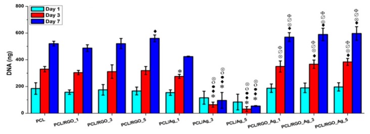
Aliphatic polyesters such as poly(lactic acid) (PLA), polycaprolactone (PCL) and poly(lactic-co-glycolic) acid (PLGA) copolymers have been widely used as biomaterials for tissue engineering applications including: bone fixation devices, bone scaffolds, and wound dressings in orthopedics. However, biodegradable aliphatic polyesters are prone to bacterial infections due to the lack of antibacterial moieties in their macromolecular chains. In this respect, silver nanoparticles (AgNPs), graphene oxide (GO) sheets and AgNPs-GO hybrids can be used as reinforcing nanofillers for aliphatic polyesters in forming antimicrobial nanocomposites. However, polymeric matrix materials immobilize nanofillers to a large extent so that they cannot penetrate bacterial membrane into cytoplasm as in the case of colloidal nanoparticles or nanosheets. Accordingly, loaded GO sheets of aliphatic polyester nanocomposites have lost their antibacterial functions such as nanoknife cutting, blanket wrapping and membrane phospholipid extraction. In contrast, AgNPs fillers of polyester nanocomposites can release silver ions for destroying bacterial cells. Thus, AgNPs fillers are more effective than loaded GO sheets of polyester nanocomposiites in inhibiting bacterial infections. Aliphatic polyester nanocomposites with AgNPs and AgNPs-GO fillers are effective to kill multi-drug resistant bacteria that cause medical device-related infections.

Keywords: Escherichia coli; aliphatic polyester; antibacterial activity; cellular viability; electrospinning; graphene oxide; osteoblast; scaffold; silver nanoparticle; staphylococcus aureus.


Conflict of interest statement

The authors declare no conflict of interest.

Figures

Figure 1
Figure 1
Bactericidal mechanisms of silver nanoparticles (AgNPs). (A) The local enlarged view shows the initial adhesion of AgNPs to the bacterial wall, leading to membrane destruction and cellular content leakage. AgNPs or silver ion (Ag+) can bind to the protein of cell membrane, thus inducing reactive oxygen species (ROS) generation and reducing adenosine triphosphate (ATP) production. (B) Penetration of AgNPs into cytoplasm; AgNPs and the released Ag+ can interact with proteins, enzymes, lipids, and deoxyribonucleic acid (DNA). The increased ROS levels lead to an apoptosis-like response, lipid peroxidation, and DNA damage. (C) AgNPs can sustainably release Ag+ inside and outside bacterial membrane, so Ag+ can interact with the proteins and enzymes accordingly.
Figure 2
Figure 2
Mechanisms of the antimicrobial activities of graphene-based materials. Reprinted with permission from [57]. Copyright American Chemical Society, 2016.
Figure 3
Figure 3
Transmission electron micrograph of GO-AgNPs (left panel) and the size distribution of AgNPs (right panel). The average size of AgNPs is 7.5 nm. Reproduced with permission from [72]. Copyright Elsevier, 2014.
Figure 4
Figure 4
(a) Time-kill curves for P. aeruginosa at different concentrations of colloidal GO-AgNPs. The error bars represent the standard deviation of experiments performed in triplicates (n = 3). Reproduced with permission from [72], Copyright Elsevier, 2014. (b) Cell viability, (c) ROS level and (d) LDH leakage of thin foils coated with AgNPs, GO and GO-AgNPs upon exposure to E. coli, S. aureus, S. epidermidis and C. albicans; C is the control foil without nanoparticles. The columns with different letters (a–d) indicate significant differences between the foil samples exposed to bacterial strains and yeast (p = 0.001); error bars are standard deviations. Reproduced with permission from [137]. Copyright Springer Open, 2018.
Figure 4
Figure 4
(a) Time-kill curves for P. aeruginosa at different concentrations of colloidal GO-AgNPs. The error bars represent the standard deviation of experiments performed in triplicates (n = 3). Reproduced with permission from [72], Copyright Elsevier, 2014. (b) Cell viability, (c) ROS level and (d) LDH leakage of thin foils coated with AgNPs, GO and GO-AgNPs upon exposure to E. coli, S. aureus, S. epidermidis and C. albicans; C is the control foil without nanoparticles. The columns with different letters (a–d) indicate significant differences between the foil samples exposed to bacterial strains and yeast (p = 0.001); error bars are standard deviations. Reproduced with permission from [137]. Copyright Springer Open, 2018.
Figure 5
Figure 5
Schematic diagram of the pressurized gyration system for fabricating polymer fibers. Reproduced with permission from [44]. Copyright Royal Society, 2018.
Figure 6
Figure 6
Schematic for the preparation strategy of PLA/AgNPs nanocomposite film.
Figure 6
Figure 6
Schematic for the preparation strategy of PLA/AgNPs nanocomposite film.
Figure 7
Figure 7
Bacterial population in colony forming unit (UFC/mL) of different S. aureus strains and P. aeruginosa strains adhered on P0, PI and P2 films. *, ** and *** correspond to the magnitude of antimicrobial activity. MRSA: Methicillin-resistant Staphylococcus aureus, and ATCC: American Type Culture Collection.
Figure 8
Figure 8
(a) Viability of osteoblasts cultured on PLA and its nanocomposites for 1, 2 and 4 days; error bars denoted standard deviations. (b) Released Ag+ ion vs. time plots of PLA/18 wt% nHA–AgNPs hybrids immersed in distilled water at 37 °C. The concentration of silver ions was determined with inductively coupled plasma atomic emission spectrometry. Reproduced with permission from [154], Copyright Royal Society of Chemistry, 2015.
Figure 9
Figure 9
Macrographs of agar plates showing bacterial inhibition zones against E. coli: (a) control (AgNO3), PLA, PLA/18 wt% nHA, PLA/18 wt% nHA–10 wt% AgNPs, PLA/18 wt% nHA–18 wt% AgNPs and PLA/18 wt% nHA–25 wt% AgNPs samples, and (b) PLA/18 wt% nHA–2 wt% AgNPs and PLA/18 wt% nHA–6 wt% AgNPs hybrid nanocomposites. Reproduced from [154], Copyright Royal Society of Chemistry, 2015.
Figure 10
Figure 10
Macrographs of agar plates showing bacterial inhibition zones against S. aureus: (a) control (AgNO3), PLA, PLA/18 wt% nHA, PLA/18 wt% nHA–10 wt% AgNPs, PLA/18 wt% nHA–18 wt% AgNPs and PLA/18 wt% nHA–25 wt% AgNPs samples, and (b) PLA/18 wt% nHA–2 wt% AgNPs and PLA/18 wt% nHA–6 wt% AgNPs hybrid nanocomposites. Reproduced with permission from [154], Copyright Royal Society of Chemistry, 2015.
Figure 11
Figure 11
SEM images of electrospun (a) PLA, (b) PLA/1 wt% GO, (c) PLA/3 wt% Ag, and (d) PLA/1 wt% GO-3 wt% Ag fibrous mats. Reproduced from [165], Copyright American Chemical Society, 2017.
Figure 12
Figure 12
(a) Images of water contact angles on PLA and its nanocomposite fibrous mats, and (b) released Ag+ concentration vs. immersion time profiles of PLA/AgNPs and PLA/1 wt%GO-AgNPs mats immersed in distilled water for different time points. The concentration of silver ions was determined with inductively coupled plasma atomic emission spectrometry. Reproduced with permission from [165], Copyright American Chemical Society, 2017.
Figure 13
Figure 13
(a) Bacterial reduction percentage vs. time profiles for E. coli culture medium treated with PLA and its nanocomposite fibrous mats. The circular disk samples were soaked in the test tubes containing nutrient broth for E. coli (1 × 10⁶ CFU/mL), and then placed in a rotary shaker at 37 °C for different time points. (b) Photograph of E. coli culture suspensions treated with PLA and its nanocomposite fibrous mats for 6 h. SEM images showing progressive loss of membrane integrity of E. coli attached on PLA/1 wt%GO-3 wt%AgNPs fibers for (c) 2, (d) 4, and (e) 6 h. White arrows indicate ruptured cell membranes. Reproduced with permission from [165], Copyright American Chemical Society, 2017.
Figure 14
Figure 14
Bacterial reduction percentage vs. time profiles for S. aureus culture medium inoculated with electrospun PLA and its nanocomposite mats. Reproduced with permission from [165], Copyright American Chemical Society, 2017.
Figure 15
Figure 15
ROS levels in (a) E. coli and (b) S. aureus treated with electrospun PLA (control) and its nanocomposite mats. * denotes statistically significant difference between the test groups (* p < 0.05). Reprinted with permission from [165], Copyright American Chemical Society, 2017.
Figure 16
Figure 16
Antibacterial activity of dense PLGA and its PLGA/AgNPs nanocomposite films against (A) S. aureus and (B) E. coli incubated for 3 and 24 h at 37 °C. The percentage viability was determined by setting the bacterial cells grown onto the tissue culture plate (TCP) wells to 100%. Error bars indicated standard errors of the means, and ** denoted statistical significance, p < 0.01.
Figure 17
Figure 17
MTT test results showing cell viability of (A) murine fibroblasts (L929) and (B) human osteosarcoma cell line (Saos-2) cultured on dense PLGA and its nanocomposite films for 24 h, 96 h and 240 h. The error bars were standard deviations.
Figure 18
Figure 18
Bacterial growth vs. exposure time profiles of neat PLGA (star), PLGA/1 wt% AgNPs (square), and PLGA/3 wt% AgNPs (circle). Reproduced with permission from [170], Copyright Wiley, 2013.
Figure 19
Figure 19
Antibacterial activity of electrospun PLGA 50/50 and PLGA 75/25 mats with and without AgNPs upon exposure to P. aeruginosa. Reproduced with permission from [172], Copyright Wiley, 2015.
Figure 20
Figure 20
Proliferation of human dermal fibroblasts (HDFs) determined by AlamarBlue test. * Significant against cell proliferation on PLGA 50/50–6%AgNPs scaffold at p ≤ 0.05; # Significant against cell proliferation on PLGA75/25–6%AgNPs scaffold at p ≤ 0.05. NP: AgNPs. Reproduced with permission from [172], Copyright Wiley, 2015.
Figure 21
Figure 21
(Top Row) Number of attached live cells on PLGA-chitosan (green column) and PLGA-chitosan/GO-AgNPs (red column) mats upon exposure to E. coli, P. aeruginosa, and S. aureus bacteria. (Bottom Row) SEM images of E. coli (A,D), P. aeruginosa (B,E), and S. aureus (C,F) attached on the PLGA-CS (AC) and PLGA-CS/GO-AgNPs (DF) mats. * denotes statistically significant difference between PLGA-CS/GO-AgNPs and PLGA-CS (control); p < 0.05. Reproduced with permission from [173], Copyright American Chemical Society, 2015.
Figure 21
Figure 21
(Top Row) Number of attached live cells on PLGA-chitosan (green column) and PLGA-chitosan/GO-AgNPs (red column) mats upon exposure to E. coli, P. aeruginosa, and S. aureus bacteria. (Bottom Row) SEM images of E. coli (A,D), P. aeruginosa (B,E), and S. aureus (C,F) attached on the PLGA-CS (AC) and PLGA-CS/GO-AgNPs (DF) mats. * denotes statistically significant difference between PLGA-CS/GO-AgNPs and PLGA-CS (control); p < 0.05. Reproduced with permission from [173], Copyright American Chemical Society, 2015.
Figure 22
Figure 22
(Top Row) Number of attached live cells on PLGA-chitosan (green column) and PLGA-chitosan/GO (red column) mats upon exposure to E. coli, P. aeruginosa, and S. aureus bacteria. (Bottom Row) images of E. coli (A), P. aeruginosa (B), and S. aureus (C) attached on the PLGA-CS/GO samples. Reproduced with permission from [173], Copyright American Chemical Society, 2015.
Figure 23
Figure 23
Images of S1, S2, S3, S4, S5 and S6 coated PCL mats showing (a) inhibition zones (b) zone diameters against S. aureus and P. aeruginosa. Reproduced with permission from [149]. Copyright Elsevier, 2018.
Figure 23
Figure 23
Images of S1, S2, S3, S4, S5 and S6 coated PCL mats showing (a) inhibition zones (b) zone diameters against S. aureus and P. aeruginosa. Reproduced with permission from [149]. Copyright Elsevier, 2018.
Figure 24
Figure 24
Photographs showing the contraction of dorsal wounds treated with S1 mat (top panel), and S6 mat (bottom panel) at day 0, day 7 and day 10, respectively. Reproduced with permission from [149]. Copyright Elsevier, 2018.
Figure 25
Figure 25
(A) SEM images of biofilms formed on PCL, PCL/DA, PCL/NS0.5, PCL/NS1.0 and PCL/NS2.0 mat surfaces; Scale bars: 1 μm. Red arrows indicate adherent bacteria. Measured areas covered by the biofilms of (B) S. aureus, (C) E. coli and (D) A. baumannii. ** p < 0.01. Reproduced with permission from [183], Dove Medical Press Limited under the Creative Commons Attribution—Non-Commercial (unported, v3.0) License.
Figure 26
Figure 26
(A) Representative macroscopic images of mice wounds from the blank, control, PCL, PCL/DA and PCL/NS1.0 groups. (B) In vivo quantitative wound closure in mice at different time points. Data are presented as mean ± SD (n = 5). Significant differences exist between PCL/NS1.0 and the control, as well as PCL and PCL/DA groups, * p < 0.05, ** p < 0.01.
Figure 27
Figure 27
Antibacterial rates of PCL and AgNPs-loaded PCL fibrous mats prepared by pressurized melt gyration against (a) E. coli and (b) P. aeruginosa after 2 h of exposure. Reproduced with permission from [39]. Copyright Wiley-VCH, 2016.
Figure 28
Figure 28
Colony count on composite surfaces incubated with E. coli for 3 h with respect to neat PCL. Statistically significant differences (p < 0.05) compared to PCL, PCL/RGO_1, PCL/RGO_3, PCL/RGO_5, PCL/Ag_1 and PCL/Ag_3 and PCL/Ag_5, PCL/RGO_Ag_1 are indicated by *, ♦, •, ο, ⊗, ∅, Φ and ⊕, respectively. Reproduced with permission from [187], Copyright Elsevier, 2016.
Figure 29
Figure 29
Released silver ions from PCL/Ag and PCL/RGO-Ag composite systems in pure water at 37 °C. Inset shows the enlarged view for values for PCL/ RGO-Ag system. Statistically significant differences (p < 0.05) compared to PCL/Ag_1 and PCL/Ag_3 and PCL/Ag_5 and PCL/RGO_Ag_1 are indicated by ⊗, ∅, Φ and ♢, respectively. Reproduced with permission from [187], Copyright Elsevier, 2016.
Figure 30
Figure 30
DNA quantification of hMSCs cultured on PCL and its composites for 1, 3 and 7 days. Statistically significant differences (p < 0.05) compared to PCL, PCL/RGO_1, PCL/RGO_3, PCL/RGO_5, PCL/Ag_1 and PCL/Ag_3 and PCL/Ag_5 are indicated by *, ♦, •, ο, ⊗, ∅ and Φ, respectively. Reproduced with permission from [187], Copyright Elsevier, 2016.

References

    1. Polívková M., Hubáček T., Staszek M., Švorčík V., Siegel J. Antimicrobial treatment of polymeric medical devices by silver nanomaterials and related technology. Int. J. Mol. Sci. 2017;18:419. doi: 10.3390/ijms18020419. - DOI - PMC - PubMed
    1. Sánchez-Salcedo S., Colilla M., Izquierdo-Barba I., Vallet-Regi M. Preventing bacterial adhesion on scaffolds for bone tissue engineering. Int. J. Bioprint. 2016;2:20–34. doi: 10.18063/IJB.2016.01.008. - DOI
    1. Hughes H., Webber M.A. Novel approaches to the treatment of bacterial biofilm infections. Br. J. Pharmacol. 2017;174:2237–2246. doi: 10.1111/bph.13706. - DOI - PMC - PubMed
    1. Cunliffe D., Smart A.C., Alexander C., Vulfson E.N. Bacterial adhesion of synthetic surfaces. Appl. Environ. Microbiol. 1999;65:4995–5002. - PMC - PubMed
    1. Renner L.D., Weibel D.B. Physicochemical regulation of biofilm formation. MRS Bull. 2011;36:347–355. doi: 10.1557/mrs.2011.65. - DOI - PMC - PubMed

LinkOut - more resources