. 2019 Aug 1;11(8):367.
doi: 10.3390/pharmaceutics11080367.

Improved In Vitro Model for Intranasal Mucosal Drug Delivery: Primary Olfactory and Respiratory Epithelial Cells Compared with the Permanent Nasal Cell Line RPMI 2650

Affiliations

Improved In Vitro Model for Intranasal Mucosal Drug Delivery: Primary Olfactory and Respiratory Epithelial Cells Compared with the Permanent Nasal Cell Line RPMI 2650

Simone Ladel et al. Pharmaceutics. .

Abstract

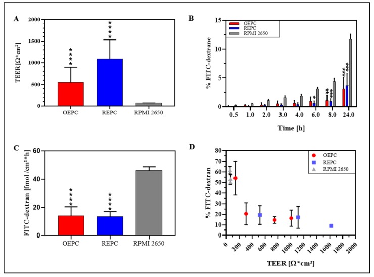
Background: The epithelial layer of the nasal mucosa is the first barrier for drug permeation during intranasal drug delivery. With increasing interest for intranasal pathways, adequate in vitro models are required. Here, porcine olfactory (OEPC) and respiratory (REPC) primary cells were characterised against the nasal tumour cell line RPMI 2650.

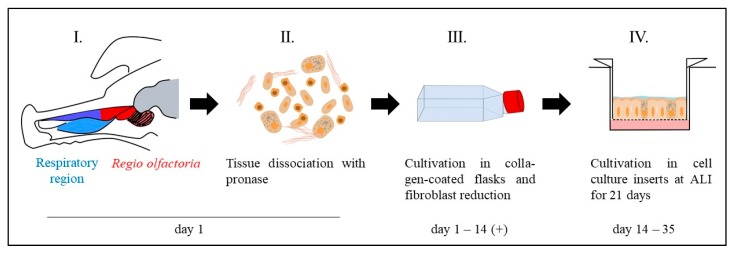
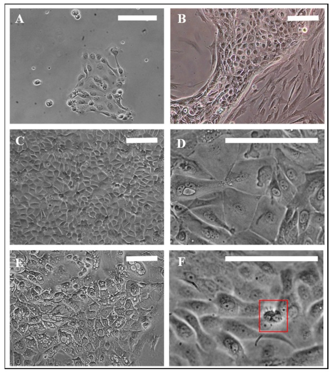
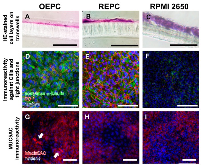
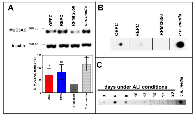
Methods: Culture conditions for primary cells from porcine nasal mucosa were optimized and the cells characterised via light microscope, RT-PCR and immunofluorescence. Epithelial barrier function was analysed via transepithelial electrical resistance (TEER), and FITC-dextran was used as model substance for transepithelial permeation. Beating cilia necessary for mucociliary clearance were studied by immunoreactivity against acetylated tubulin.

Results: OEPC and REPC barrier models differ in TEER, transepithelial permeation and MUC5AC levels. In contrast, RPMI 2650 displayed lower levels of MUC5AC, cilia markers and TEER, and higher FITC-dextran flux rates.

Conclusion: To screen pharmaceutical formulations for intranasal delivery in vitro, translational mucosal models are needed. Here, a novel and comprehensive characterisation of OEPC and REPC against RPMI 2650 is presented. The established primary models display an appropriate model for nasal mucosa with secreted MUC5AC, beating cilia and a functional epithelial barrier, which is suitable for long-term evaluation of sustained release dosage forms.

Keywords: RPMI 2650; barrier model; nose-to-brain; olfactory epithelium; primary cells; respiratory epithelium.


Conflict of interest statement

The authors declare no conflicts of interest.

Figures

Figure 1
Figure 1
Workflow for primary cell isolation and cultivation under air–liquid interface (ALI) conditions. The mucosa was dissected either from the olfactory region (red) or the respiratory region (blue) (I.). Single cells were obtained by pronase digestion of the mucosa explants (II.). Single cells were cultivated in collagen-coated T75 cell culture flasks (III.). To reduce fibroblast overgrowth, the culture was shortly trypsinated (2–4 min) to get rid of the less adherent fibroblasts and select for epithelial cells. The cells were cultivated up to 80–90% confluence in the T75 flasks and transferred into cell culture inserts to grow under ALI conditions for 21 days (IV.).
Figure 2
Figure 2
Morphology of porcine primary nasal epithelial cells. (A) Olfactory primary cells, 24 h in culture. (B) Fibroblast contamination in primary epithelial cells. (C,D) Morphological appearance of OEPC and REPC: small round or cobblestone shaped and big flat epithelial cells; cell membranes are clearly visible. (E) REPC: vesicular cells. (F) Red box: Ciliated cells can be motile or non-motile (beating frequency of >5 Hz). Cilia length was ~10 µm. OEPC: olfactory epithelial primary cells; REPC: respiratory epithelial primary cells; Scale bars: 100 µm.
Figure 3
Figure 3
Characterisation of olfactory and respiratory primary cells in comparison to the standard cell line RPMI 2650. Monolayers are necessary to evaluate transport over the epithelial layer in the nasal mucosa: 14 µm sections were made of olfactory epithelium primary cells (OEPC, A), respiratory epithelial primary cells (REPC, B) and RPMI 2650 (C) grown on a cell insert membrane for 21 days. Morphological features such as tight junctions and the formation of cilia are important influencing factors in investigations of drug permeation and clearance studies. Acetylated α-tubulin is a common marker for cilia [54]. IF double-staining of acetylated α-tubulin and the tight junction marker zonula occludens-1 (ZO-1) of ALI cultures of OEPC (D), REPC (E) and RPMI 2650 (F) after 21 days of incubation were made. An additional feature of mucosal cells is the ability to produce mucus. The marker protein mucin 5AC was used in this work, because the olfactory mucosa is to be simulated above all to investigate nose-to-brain transport. Again, IF was performed in OEPC (G), REPC (H) and RPMI 2650 (I). Scale bars: 100 µm.
Figure 4
Figure 4
Mucin MUC5AC expression and immunoreactivity in primary cells of the nasal cavity. (A) Transcription analysis (RT-PCR) of MUC5AC gene in olfactory epithelial primary cells (OEPC), respiratory epithelial primary cells (REPC), tumour cell line RPMI 2650 and the concha nasalis media (c.n. media). MUC5AC transcript signal was referenced to beta-actin transcript signal. The significance was calculated by comparison of the OEPC, REPC and RPMI 2650 data with the c.n. media transcription data using an unpaired t-test. * p < 0.05; n = 4; error bars represent mean ± SD. (B) Dot blot analysis of MUC5AC protein in lysates of OEPC, REPC, RPMI 2650 and c.n. media. All OEPC and REPC cultures shown in (A,B) were cultivated for 14 days in vitro in T flasks. (C) Immunoreactivity against MUC5AC in OEPC that were first cultured for 7 days in T flask with a minimum confluency of 70% then under ALI conditions additional 20 days. Apically secreted mucus was collected at the days indicated corresponding to a mucin production of 2 to 3 days. Statistical analysis: unpaired t-test, * p < 0.05 compared to the standard model RPMI 2650.
Figure 5
Figure 5
Comparison of FITC-dextran permeation and TEER value of nasal primary cells vs. RPMI 2650. (A) TEER values of OEPC, REPC and RPMI 2650 after 21 days ALI. (B) FITC-dextran permeation data: Values of FITC-dextran permeated through a cell layer were normalized to total of FITC-dextran. (C) Flux of FITC-dextran through an OEPC, REPC and RPMI 2650 layer after 24 h. (D) Correlation of percentage FITC-dextran permeation after 24 h and TEER value. Statistical analysis: unpaired t-test, * p < 0.05, ** p < 0.01, *** p < 0.001, **** p < 0.0001; all compared to the standard model RPMI 2650. OEPC: olfactory epithelial primary cells; REPC: respiratory epithelial primary cells; TEER: Transepithelial electrical resistance. FITC-dextran: Fluorescein isothiocyanate-dextran; N = 4, n = 21; error bars represent mean ± SD of all repetitions.

Similar articles

Cited by

References

    1. Barnabas W. Drug targeting strategies into the brain for treating neurological diseases. J. Neurosci. Methods. 2019;311:133–146. doi: 10.1016/j.jneumeth.2018.10.015. - DOI - PubMed
    1. Illum L. Nasal drug delivery: New developments and strategies. Drug Discov. Today. 2002;7:1184–1189. doi: 10.1016/S1359-6446(02)02529-1. - DOI - PubMed
    1. Neuwelt E., Abbott N.J., Abrey L., Banks W.A., Blakley B., Engelhardt B., Grammas P., Nedergaard M., Nutt J., Pardridge W., et al. Strategies to advance translational research into brain barriers. Lancet Neurol. 2008;7:84–96. doi: 10.1016/S1474-4422(07)70326-5. - DOI - PubMed
    1. Gao H. Progress and perspectives on targeting nanoparticles for brain drug delivery. Acta Pharm. Sin. B. 2016;6:268–286. doi: 10.1016/j.apsb.2016.05.013. - DOI - PMC - PubMed
    1. Illum L. Nasal drug delivery—Possibilities, problems and solutions. J. Control. Release. 2003;87:187–198. doi: 10.1016/S0168-3659(02)00363-2. - DOI - PubMed

LinkOut - more resources