

Stereoselective Anti-Cancer Activities of Ginsenoside Rg3 on Triple Negative Breast Cancer Cell Models
- PMID: 31374984
- PMCID: PMC6789838
- DOI: 10.3390/ph12030117
Stereoselective Anti-Cancer Activities of Ginsenoside Rg3 on Triple Negative Breast Cancer Cell Models
Abstract
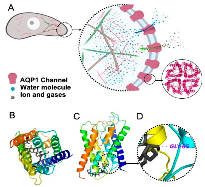
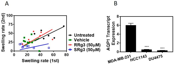
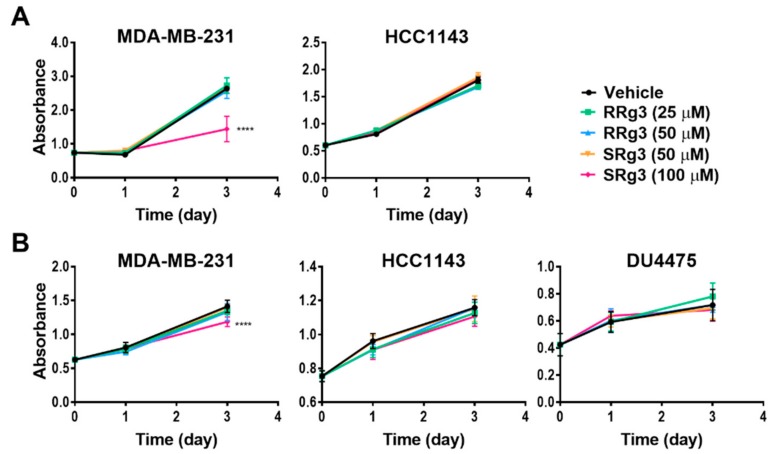
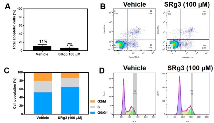
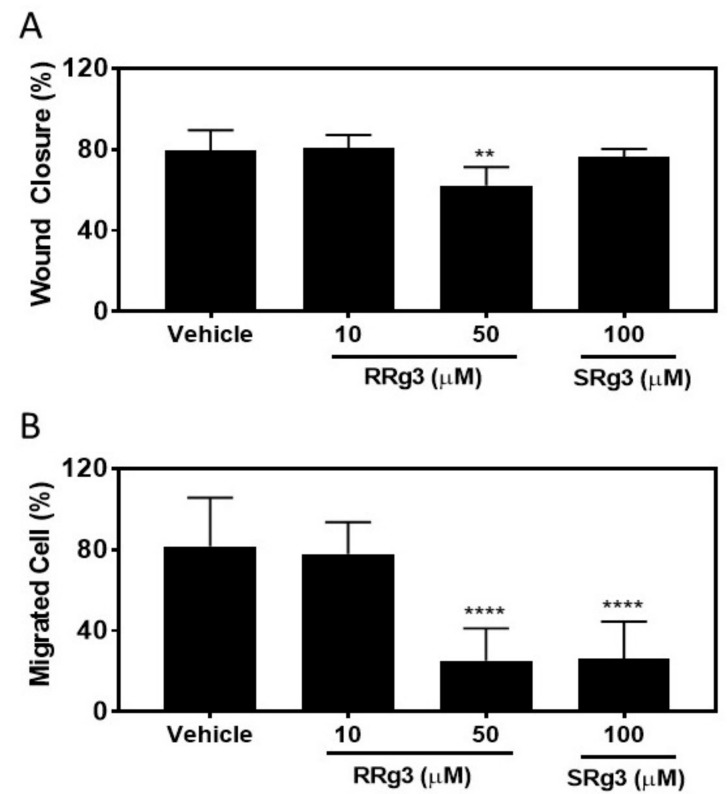
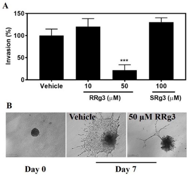
Ginsenoside Rg3 (Rg3) has two epimers, 20(S)-ginsenoside Rg3 (SRg3) and 20(R)-ginsenoside Rg3 (RRg3), and while Rg3 itself has been reported to have anti-cancer properties, few studies have been reported on the anti-cancer effects of the different epimers. The aim was to investigate the stereoselective effects of the Rg3 epimers on triple negative breast cancer (TNBC) cell lines, tested using cell-based assays for proliferation, apoptosis, cell cycle arrest, migration and invasion. Molecular docking showed that Rg3 interacted with the aquaporin 1 (AQP1) water channel (binding score -9.4 kJ mol-1). The Xenopus laevis oocyte expression system was used to study the effect of Rg3 epimers on the AQP1 water permeability. The AQP1 expression in TNBC cell lines was compared with quantitative-polymerase chain reaction (PCR). The results showed that only SRg3 inhibited the AQP1 water flux and inhibited the proliferation of MDA-MB-231 (100 μM), due to cell cycle arrest at G0/G1. SRg3 inhibited the chemoattractant-induced migration of MDA-MB-231. The AQP1 expression in MDA-MB-231 was higher than in HCC1143 or DU4475 cell lines. These results suggest a role for AQP1 in the proliferation and chemoattractant-induced migration of this cell line. Compared to SRg3, RRg3 had more potency and efficacy, inhibiting the migration and invasion of MDA-MB-231. Rg3 has stereoselective anti-cancer effects in the AQP1 high-expressing cell line MDA-MB-231.
Keywords: Ginsenoside Rg3; breast cancer; epimer; stereoselective; triple negative breast cancer.
Conflict of interest statement
The authors declare no conflict of interest.
Figures
Similar articles
-
Anti-Cancer Effects of an Optimised Combination of Ginsenoside Rg3 Epimers on Triple Negative Breast Cancer Models.Pharmaceuticals (Basel). 2021 Jun 30;14(7):633. doi: 10.3390/ph14070633. Pharmaceuticals (Basel). 2021. PMID: 34208799 Free PMC article.
-
Anti-Angiogenic Properties of Ginsenoside Rg3 Epimers: In Vitro Assessment of Single and Combination Treatments.Cancers (Basel). 2021 May 6;13(9):2223. doi: 10.3390/cancers13092223. Cancers (Basel). 2021. PMID: 34066403 Free PMC article.
-
Differential antiangiogenic and anticancer activities of the active metabolites of ginsenoside Rg3.J Ginseng Res. 2024 Mar;48(2):171-180. doi: 10.1016/j.jgr.2021.05.008. Epub 2021 Jun 3. J Ginseng Res. 2024. PMID: 38465222 Free PMC article.
-
Ginsenoside Rg3 promotes cytotoxicity of Paclitaxel through inhibiting NF-κB signaling and regulating Bax/Bcl-2 expression on triple-negative breast cancer.Biomed Pharmacother. 2017 May;89:227-232. doi: 10.1016/j.biopha.2017.02.038. Epub 2017 Feb 20. Biomed Pharmacother. 2017. PMID: 28231544
-
Anti-Angiogenic Properties of Ginsenoside Rg3.Molecules. 2020 Oct 23;25(21):4905. doi: 10.3390/molecules25214905. Molecules. 2020. PMID: 33113992 Free PMC article. Review.
Cited by
-
Bactericidal and In Vitro Cytotoxicity of Moringa oleifera Seed Extract and Its Elemental Analysis Using Laser-Induced Breakdown Spectroscopy.Pharmaceuticals (Basel). 2020 Aug 13;13(8):193. doi: 10.3390/ph13080193. Pharmaceuticals (Basel). 2020. PMID: 32823699 Free PMC article.
-
Network Pharmacology-Based Approach to Investigate the Molecular Targets of Sinomenine for Treating Breast Cancer.Cancer Manag Res. 2021 Feb 9;13:1189-1204. doi: 10.2147/CMAR.S282684. eCollection 2021. Cancer Manag Res. 2021. PMID: 33603465 Free PMC article.
-
Natural Products as Novel Therapeutic Agents for Triple-Negative Breast Cancer: Current Evidence, Mechanisms, Challenges, and Opportunities.Molecules. 2025 Mar 7;30(6):1201. doi: 10.3390/molecules30061201. Molecules. 2025. PMID: 40141978 Free PMC article. Review.
-
Effect of Zebularine on p16INK4a, p14ARF, p15INK4b, and DNA Methyltransferase 1 Gene Expression, Cell Growth Inhibition, and Apoptosis Induction in Human Hepatocellular Carcinoma PLC/PRF5 and Pancreatic Cancer PA-TU-8902 Cell Lines.Iran J Pharm Res. 2020 Fall;19(4):193-202. doi: 10.22037/ijpr.2020.112223.13614. Iran J Pharm Res. 2020. PMID: 33841535 Free PMC article.
-
Anticancer Activities of Ginsenosides, the Main Active Components of Ginseng.Evid Based Complement Alternat Med. 2021 Feb 3;2021:8858006. doi: 10.1155/2021/8858006. eCollection 2021. Evid Based Complement Alternat Med. 2021. PMID: 33623532 Free PMC article. Review.
References
-
- Yang M.S., Wu M.Y. Nutraceuticals. Elsevier; Amsterdam, The Nederlands: 2016. Chinese ginseng; pp. 693–705.
Grants and funding
LinkOut - more resources
Full Text Sources
Miscellaneous
