. 2019 Aug 1;10(3):35.
doi: 10.3390/jfb10030035.

Biocompatibility of Nanocellulose-Reinforced PVA Hydrogel with Human Corneal Epithelial Cells for Ophthalmic Applications

Affiliations

Biocompatibility of Nanocellulose-Reinforced PVA Hydrogel with Human Corneal Epithelial Cells for Ophthalmic Applications

Gopi Krishna Tummala et al. J Funct Biomater. .

Abstract

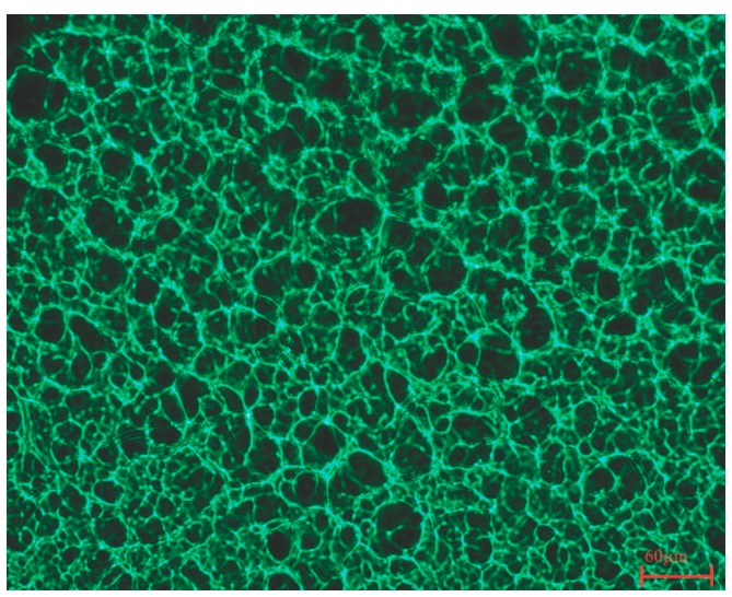
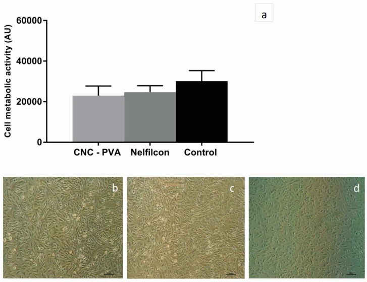
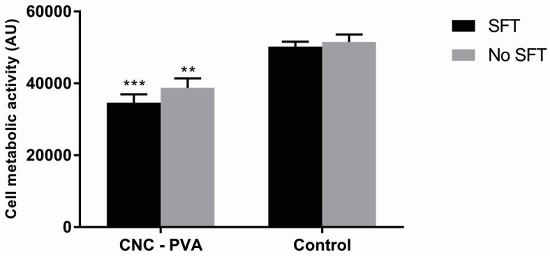
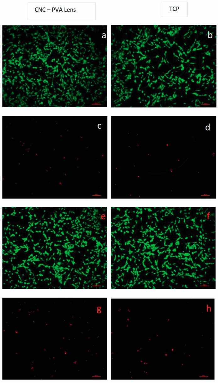
Transparent composite hydrogel in the form of a contact lens made from poly(vinyl alcohol) (PVA) and cellulose nanocrystals (CNCs) was subjected to in vitro biocompatibility evaluation with human corneal epithelial cells (HCE-2 cells). The cell response to direct contact with the hydrogels was investigated by placing the samples on top of confluent cell layers and evaluating cell viability, morphology, and cell layer integrity subsequent to 24 h culture and removal of the hydrogels. To further characterize the lens-cell interactions, HCE-2 cells were seeded on the hydrogels, with and without simulated tear fluid (STF) pre-conditioning, and cell viability and morphology were evaluated. Furthermore, protein adsorption on the hydrogel surface was investigated by incubating the materials with STF, followed by protein elution and quantification. The hydrogel material was found to have affinity towards protein adsorption, most probably due to the interactions between the positively charged lysozyme and the negatively charged CNCs embedded in the PVA matrix. The direct contact experiment demonstrated that the physical presence of the lenses did not affect corneal epithelial cell monolayers in terms of integrity nor cell metabolic activity. Moreover, it was found that viable corneal cells adhered to the hydrogel, showing the typical morphology of epithelial cells and that such response was not influenced by the STF pre-conditioning of the hydrogel surface. The results of the study confirm that PVA-CNC hydrogel is a promising ophthalmic biomaterial, motivating future in vitro and in vivo biocompatibility studies.

Keywords: cellulose nanocrystals; contact lens; cornea regeneration; poly(vinyl alcohol); therapeutic lens.


Conflict of interest statement

The authors declare no conflict of interest.

Figures

Figure 1
Figure 1
Cross section image of cellulose nanocrystals (CNC)-poly(vinyl alcohol) (PVA) hydrogel acquired with an IF 550 green contrast color filter. Scale bar corresponds to 60 µm.
Figure 2
Figure 2
Human corneal epithelial cells (HCE-2 cells) after 24 h direct contact with the hydrogels (CNC-PVA and Nelfilcon) and subsequent removal of the materials. (a) Cell metabolic activity assessed by the Alamar blue assay. Data represent the mean ± standard error of the mean (SEM) for n = 3. One way ANOVA was applied. No significant difference was observed between cells exposed to the hydrogels and the control (untreated cells); (bd) representative light microscopy images of the cell layers after direct contact with (b) CNC-PVA and (c) Nelfilcon A; while (d) corresponds to untreated cells. The scale bar corresponds to 100 µm.
Figure 3
Figure 3
Cell viability of HCE-2 cells cultured on CNC-PVA lens and tissue culture plate (control) for 24 h, with and without simulated tear fluid (STF) pre-conditioning of the material surfaces (SFT and no SFT, respectively). Data represent the mean ± SEM for n = 3. Statistically significant differences between the CNC-PVA and the controls are indicated with ** p < 0.01 and *** p < 0.001 by two-way ANOVA.
Figure 4
Figure 4
Live/dead staining of HCE-2 cells cultured on CNC-PVA lens and tissue culture plate (TCP) for 24 h. Panels (a,b) and (c,d) show images of live (green) and dead cells (red), respectively, in surfaces pre-conditioned with STF, while panels (e,f) and (g,h) show images of live (green) and dead cells (red), respectively, in materials not previously exposed to STF. The scale bar corresponds to 100 µm.

References

    1. Sridhar M.S. Anatomy of cornea and ocular surface. Indian J. Ophthalmol. 2018;66:190–194. - PMC - PubMed
    1. Lloyd A., Faragher R., Wassall M., Rhys-Williams W., Wong L., Hughes J., Hanlon G. Assessing the in vitro cell based ocular compatibility of contact lens materials. Contact Lens Anter. Eye. 2000;23:119–123. doi: 10.1016/S1367-0484(00)80004-1. - DOI - PubMed
    1. Sakata M., Sack R.A., Sathe S., Holden B., Beaton A.R. Polymorphonuclear leukocyte cells and elastase in tears. Curr. Eye Res. 1997;16:810–819. doi: 10.1076/ceyr.16.8.810.8992. - DOI - PubMed
    1. Oliva M.S., Schottman T., Gulati M. Turning the tide of corneal blindness. Indian J. Ophthalmol. 2012;60:423. doi: 10.4103/0301-4738.100540. - DOI - PMC - PubMed
    1. Myung D., Duhamel P.E., Cochran J.R., Noolandi J., Ta C.N., Frank C.W. Development of Hydrogel-Based Keratoprostheses: A Materials Perspective. Biotechnol. Prog. 2008;24:735–741. doi: 10.1021/bp070476n. - DOI - PMC - PubMed

LinkOut - more resources