Review
. 2019 Aug 2;14(1):32.
doi: 10.1186/s13024-019-0333-5.

The neuropathological diagnosis of Alzheimer's disease

Affiliations
Review

The neuropathological diagnosis of Alzheimer's disease

Michael A DeTure et al. Mol Neurodegener. .

Abstract

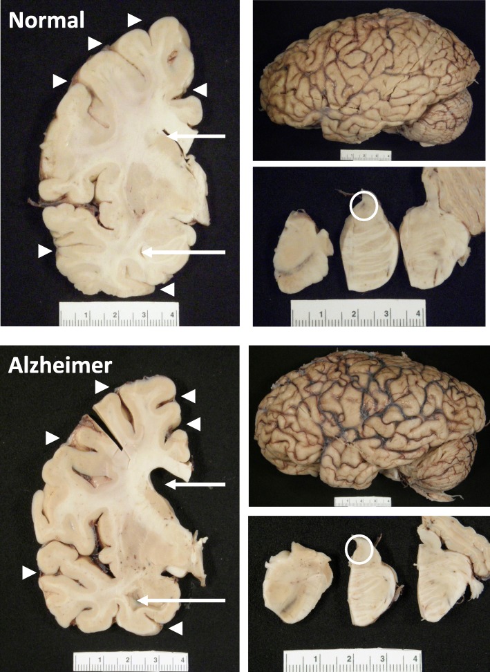
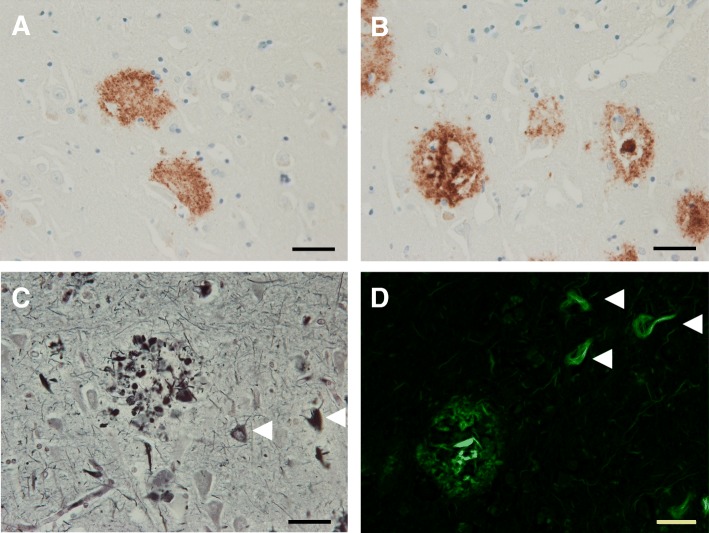
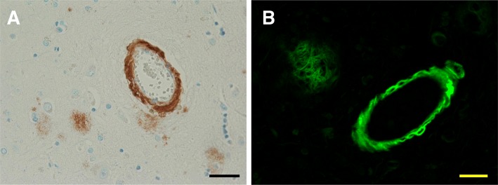
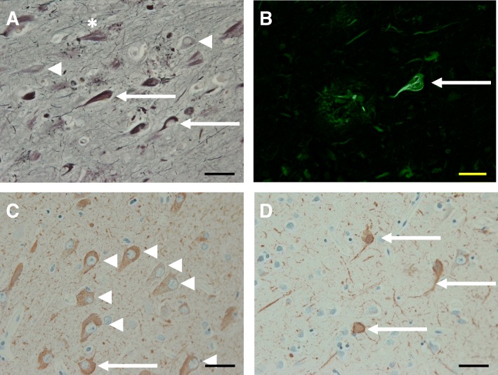
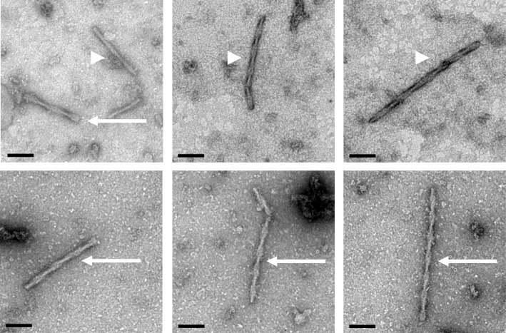
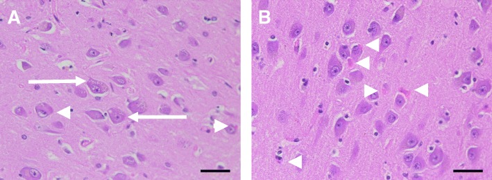
Alzheimer's disease is a progressive neurodegenerative disease most often associated with memory deficits and cognitive decline, although less common clinical presentations are increasingly recognized. The cardinal pathological features of the disease have been known for more than one hundred years, and today the presence of these amyloid plaques and neurofibrillary tangles are still required for a pathological diagnosis. Alzheimer's disease is the most common cause of dementia globally. There remain no effective treatment options for the great majority of patients, and the primary causes of the disease are unknown except in a small number of familial cases driven by genetic mutations. Confounding efforts to develop effective diagnostic tools and disease-modifying therapies is the realization that Alzheimer's disease is a mixed proteinopathy (amyloid and tau) frequently associated with other age-related processes such as cerebrovascular disease and Lewy body disease. Defining the relationships between and interdependence of various co-pathologies remains an active area of investigation. This review outlines etiologically-linked pathologic features of Alzheimer's disease, as well as those that are inevitable findings of uncertain significance, such as granulovacuolar degeneration and Hirano bodies. Other disease processes that are frequent, but not inevitable, are also discussed, including pathologic processes that can clinically mimic Alzheimer's disease. These include cerebrovascular disease, Lewy body disease, TDP-43 proteinopathies and argyrophilic grain disease. The purpose of this review is to provide an overview of Alzheimer's disease pathology, its defining pathologic substrates and the related pathologies that can affect diagnosis and treatment.

Keywords: Alzheimer’s disease; Amyloid plaques; Neurofibrillary tangles; Neuropathology.


Conflict of interest statement

The authors declare that they have no competing interests.

Figures

Fig. 1
Fig. 1
Gross Anatomy of Alzheimer’s Brain. Lateral view of an Alzheimer’s brain can show widening of sulcal spaces and narrowing of gyri compared to a normal brain. This may be more readily observed in coronal sections as indicated by the arrowheads, and this atrophy is often accompanied by enlargement of the frontal and temporal horns of the lateral ventricles as highlighted by the arrows. Additionally, loss of pigmented neurons in the locus coeruleus is commonly observed in the pontine tegmentum as shown with the open circle. None of these features is exclusive to Alzheimer’s disease
Fig. 2
Fig. 2
Alzheimer Senile Plaques. Immunohistochemistry of affected Alzheimer’s tissues using antibodies directed against Aβ peptides demonstrates the presence of both diffuse (a) and dense core (b) senile plaques. These dense core plaques are often associated with neuritic elements that can stain filamentous tau and correlate with disease severity. Neuritic AD plaques are readily observed using Bielchowsky silver staining (c) or Thioflavin S staining (d). These stains can also label neurofibrillary tangles as shown by the arrowheads. The scale bars are 40 μm
Fig. 3
Fig. 3
Cerebral Amyloid Angiopathy. Cerebral amyloid angiopathy or congophilic amyloid angiopathy can by visualized in frontal cortical sections using Aβ directed immunohistochemistry (a) or Thioflavin S staining (b) similar to that used to detect senile plaques, and they are believed to contribute independently to the Alzheimer’s disease course. Scale bars are 40 μm
Fig. 4
Fig. 4
Neurofibrillary Tangles. Neurofibrillary tangles develop from intracellular pre-tangles containing misfolded tau and small tau aggregates to mature NFTs containing bundles of cross-linked tau filaments before the neuron dies and an extracellular ghost tangle (asterisk) remains. Silver staining (a) and Thioflavin S (b) capture many mature tangles (arrows) and some pre-tangles (arrowheads) along with amyloid plaques and tau neuropil threads. Development of NFTS from the pre-tangles is more easily visualized using tau immunohistochemistry (c, d). This allows the mis-localized somal tau to be distinguished readily from the bundles of PHFs in NFTS in addition to the neuropil threads that can also be pronounced (d). The scale bars are 40 μm
Fig. 5
Fig. 5
Paired Helical Filaments and Straight Filaments. Neurofibrillary tangles are composed of insoluble tau filaments that can be extracted using detergents or acids and visualized using electron microscopy and negative staining such as 2% uranyl acetate here. The paired helical filaments (arrows) and straight filaments (arrowheads) both appear to be composed of pairs of 10 nm filaments that wrap around one another with the PHFs exhibiting wider 10–20 nm modulations in diameter and the SFs narrower 10–15 nm modulations. Length bars are 100 nm
Fig. 6
Fig. 6
Granulovacuolar Degeneration and Hirano Bodies. Granulovacuolar degeneration is commonly observed in the neurons of AD (a). As indicated by the arrows, these neurons contain numerous vacuoles housing a dense granule. Additionally, Hirano bodies are often observed as eosinophilic pink rods within the neurons, as shown with the arrowheads (b). The scale bars are 40 μm

References

    1. Alzheimer A, Stelzmann RA, Schnitzlein HN, Murtagh FR. An English translation of Alzheimer's 1907 paper, "Uber eine eigenartige Erkankung der Hirnrinde". Clin Anat. 1995;8:429–431. doi: 10.1002/ca.980080612. - DOI - PubMed
    1. Schultz C., Del Tredici K.H.B: Neuropathology of Alzheimer’s Disease. In Alzheimer’s Disease Current Clinical Neurology. Edited by R. R, B. R. Totowa, NJ: Humana Press; 2004.
    1. Murray ME, Graff-Radford NR, Ross OA, Petersen RC, Duara R, Dickson DW. Neuropathologically defined subtypes of Alzheimer's disease with distinct clinical characteristics: a retrospective study. Lancet Neurol. 2011;10:785–796. doi: 10.1016/S1474-4422(11)70156-9. - DOI - PMC - PubMed
    1. Caselli RJ, Beach TG, Knopman DS, Graff-Radford NR. Alzheimer disease: scientific breakthroughs and translational challenges. Mayo Clin Proc. 2017;92:978–994. doi: 10.1016/j.mayocp.2017.02.011. - DOI - PMC - PubMed
    1. Aisen PS, Cummings J, Jack CR, Jr, Morris JC, Sperling R, Frolich L, Jones RW, Dowsett SA, Matthews BR, Raskin J, et al. On the path to 2025: understanding the Alzheimer's disease continuum. Alzheimers Res Ther. 2017;9:60. doi: 10.1186/s13195-017-0283-5. - DOI - PMC - PubMed

Publication types

MeSH terms