. 2019 Oct 4;294(40):14574-14590.
doi: 10.1074/jbc.RA119.009200. Epub 2019 Aug 2.

Structural and functional analysis of parameters governing tankyrase-1 interaction with telomeric repeat-binding factor 1 and GDP-mannose 4,6-dehydratase

Affiliations

Structural and functional analysis of parameters governing tankyrase-1 interaction with telomeric repeat-binding factor 1 and GDP-mannose 4,6-dehydratase

Travis Eisemann et al. J Biol Chem. .

Abstract

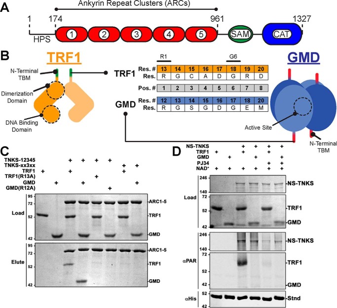
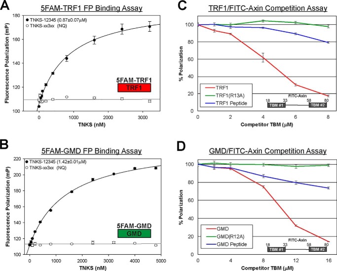
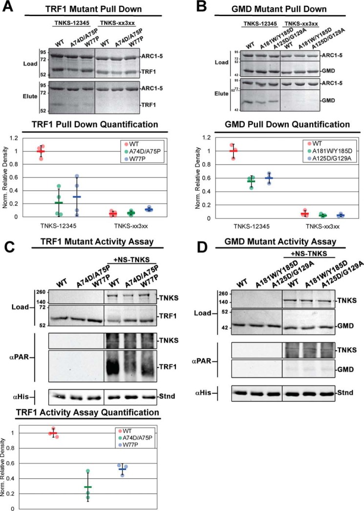
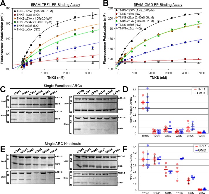
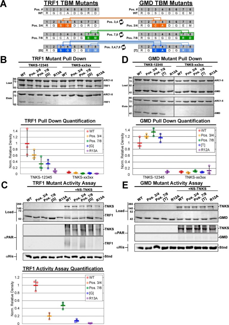
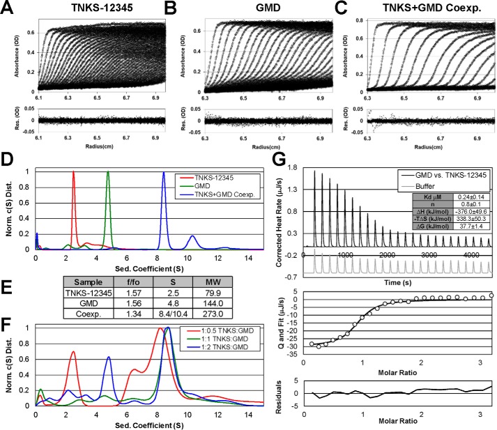
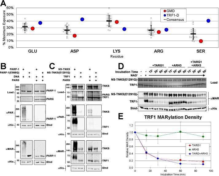
Human tankyrase-1 (TNKS) is a member of the poly(ADP-ribose) polymerase (PARP) superfamily of proteins that posttranslationally modify themselves and target proteins with ADP-ribose (termed PARylation). The TNKS ankyrin repeat domain mediates interactions with a growing number of structurally and functionally diverse binding partners, linking TNKS activity to multiple critical cell processes, including Wnt signaling, Golgi trafficking, and telomere maintenance. However, some binding partners can engage TNKS without being modified, suggesting that separate parameters influence TNKS interaction and PARylation. Here, we present an analysis of the sequence and structural features governing TNKS interactions with two model binding partners: the PARylated partner telomeric repeat-binding factor 1 (TRF1) and the non-PARylated partner GDP-mannose 4,6-dehydratase (GMD). Using a combination of TNKS-binding assays, PARP activity assays, and analytical ultracentrifugation sedimentation analysis, we found that both the specific sequence of a given TNKS-binding peptide motif and the quaternary structure of individual binding partners play important roles in TNKS interactions. We demonstrate that GMD forms stable 1:1 complexes with the TNKS ankyrin repeat domain; yet, consistent with results from previous studies, we were unable to detect GMD modification. We also report in vitro evidence that TNKS primarily directs PAR modification to glutamate/aspartate residues. Our results suggest that TNKS-binding partners possess unique sequence and structural features that control binding and PARylation. Ultimately, our findings highlight the binding partner:ankyrin repeat domain interface as a viable target for inhibition of TNKS activity.

Keywords: ADP-ribosylation; GDP-mannose dehydratase, GMD; enzyme mechanism; enzyme structure; poly(ADP-ribose) polymerase; post-translational modification (PTM); protein-protein interaction; tankyrase; telomere repeat factor 1, TRF1.


Conflict of interest statement

The authors declare that they have no conflicts of interest with the contents of this article

Figures

Figure 1.
Figure 1.
TRF1 and GMD as example TNKS-binding partners. A, schematic of TNKS domain architecture and regulatory domains. B, cartoon depictions of TRF1 and GMD quaternary structures illustrating TBMs, functional domains, and active sites. The TBM sequences (Res.) and residue numbering (Res. #) of TRF1 and GMD are compared (center). Strictly required residues are noted (R1 and G6), and the standard TBM position numbering (Pos. #) is shown (gray). C, pulldown assay utilizing histidine-tagged ARC1–5 constructs and untagged full-length TRF1 or GMD. TNKS constructs consisted of WT TNKS-12345 and quadruple ARC mutant construct TNKS-xx3xx. Binding partner constructs consisted of WT or TBM mutants R13A and R12A for TRF1 and GMD, respectively. Input proteins (Load) and elution fractions (Elute) were analyzed by SDS-PAGE. D, PARP activity assay utilizing NS-TNKS and full-length TRF1 and GMD. The designated reactions mixtures were analyzed by SDS-PAGE (Load) and by Western blot analysis of PARylation (αPAR). A His-tagged protein (Stnd) was included in each reaction to assess transfer efficiency of the blot; the protein was detected with an anti-His antibody (αHis). NS-TNKS was loaded at loaded at 0.5 μm, TRF1 and GMD were loaded at 1 μm, and PARP inhibitor PJ34 was used at 100 μm.
Figure 2.
Figure 2.
TNKS binding to TRF1 and GMD. A and B, FP binding analysis of 5FAM-TRF1 (A) and 5FAM-GMD (B) peptide interaction with TNKS-12345 (ankyrin repeat region) and TNKS-xx3xx (ankyrin repeat region with no functional ARCs). Binding was considered NQ if the binding curve did not allow for robust KD determination. Reactions that exhibited NQ binding were analyzed in at least two separate experiments, and all other KD values were determined from a minimum of three different experiments. Data represent the mean, error bars represent S.E. C and D, competition fluorescence polarization binding analysis of full-length TRF1 (C) and full-length GMD (D). A fluorescently labeled Axin peptide and TNKS-12345 were maintained at saturating concentrations (27 nm and 1.5μm, respectively) as increasing amounts of competing binding partners were added. TRF1 and GMD constructs consisted of full-length WT and TBM mutants R13A and R12A for TRF1 and GMD, respectively. For comparison, unlabeled peptides representing their TBMs were evaluated (GMD peptide and TRF1 peptide). Competitor concentrations represent the total TBM concentration. Thus, TRF1 and GMD are reported at their monomer concentrations to directly compare the relative efficiency of full-length proteins versus peptides. Data represent the mean, error bars represent S.D. mP, milli-polarization units.
Figure 3.
Figure 3.
The effects of quaternary structure alteration on TNKS binding to and PARylation of TRF1 and GMD. A and B, results and quantification of pulldown assays analyzing the binding of TNKS-12345 to multimerization-deficient TRF1 (A) and GMD (B) constructs. Histidine-tagged TNKS-12345 or TNKS-xx3xx (binding-deficient control) were used to pull down untagged TRF1 and GMD constructs with multimerization-disrupting mutations. For quantification, binding partner density was normalized to the density of TNKS-12345 in each respective reaction. C and D, TNKS PARP activity assays analyzing the PARylation of multimerization-deficient TRF1 (C) and GMD (D) constructs. The quantification of PARylation of TRF1 constructs is shown in the bottom plot of C. The designated reaction mixtures were analyzed by SDS-PAGE (Load) and by Western blot analysis of PARylation (αPAR). A His-tagged protein (Stnd) was added to each reaction after quenching to assess transfer efficiency of the blot; the protein was detected with an anti-His antibody (αHis). NS-TNKS, TRF1, and GMD were each loaded at 0.5μm for the TNKS activity assays. Data represent the mean, error bars represent S.D. The black divider lines in the center of each gel and Western blot designate where the image has been sliced for presentation. For C and D, see Fig. S3, C and D, for complete lanes for Western blots.
Figure 4.
Figure 4.
TNKS binding footprint of TRF1 and GMD. A and B, FP binding analysis of TRF1 (A) and GMD (B) peptide interaction with TNKS-12345 and TNKS-12345 constructs bearing a single functional ARC. Binding was deemed NQ if the binding curve did not allow for robust KD determination. TNKS variants that exhibited NQ binding were analyzed in at least two separate experiments, and all other KD values were determined from a minimum of three different experiments. Data represent the mean, error bars represent S.E. C and D, results and quantification of pulldown experiments analyzing the binding of untagged TRF1 and GMD to TNKS-12345 constructs with single functional ARCs. Input proteins (Load) and elution fractions (Elute) were analyzed by SDS-PAGE. E and F, results and quantification of pulldown experiment analyzing the binding of untagged TRF1 and GMD to TNKS constructs where each individual ARC has been rendered nonfunctional. Input proteins (Load) and elution fractions (Elute) were analyzed by SDS-PAGE. Binding data for WT construct TNKS-12345 and TNKS-xx3xx are used as a reference in D and F and are the collective result of multiple experiments. Binding partner density was normalized to the density of TNKS in each reaction. Data represent the mean, error bars represent S.D. mP, milli-polarization units.
Figure 5.
Figure 5.
The effect of TBM alteration on TNKS binding to and PARylation of TRF1 and GMD. A, schematic of TBM mutations wherein specific residues between the TRF1 and GMD TBMs were exchanged. B, results and quantification of pulldown experiments analyzing the binding of TRF1 constructs with TBM swapping mutants. Histidine-tagged TNKS constructs were either WT TNKS-12345 or TNKS-xx3xx. Input proteins (Load) and elution fractions (Elute) were analyzed by SDS-PAGE. Binding partner density was normalized to the density of TNKS in each reaction. Data represent the mean, error bars represent S.D. C, TNKS PARP activity assays and quantification analyzing the effect of TBM mutation on PARylation of TRF1. The designated reaction mixtures were analyzed by SDS-PAGE (Load) and by Western blot analysis of PARylation (αPAR). A His-tagged protein (Stnd) was added to each reaction after quenching to assess transfer efficiency of the blot; the protein was detected with an anti-His antibody (αHis). Data represent the mean, error bars represent S.D. D, results and quantification of pulldown experiments performed as in B except using GMD constructs with TBM mutations. E, TNKS PARP activity assay performed as in C except using GMD constructs with TBM mutations. For activity assays, NS-TNKS and GMD were loaded at 0.5μm, and TRF1 was loaded at 1μm to better resolve the subtle differences in PARylation density. The black divider lines in the center of the gel in D designate where the image has been sliced for presentation. For C and E, see Fig. S4, A and B, for the complete Western blot images.
Figure 6.
Figure 6.
Analysis of TNKS-12345 and GMD complex formation. A–C, SV-AUC analysis of TNKS-12345 (A), GMD (B), or the coexpressed complex of TNKS-12345 and GMD (C). The top panels show absorbance data (circles) and associated c(S) model fit (lines). The bottom panels show residuals for the fit. TNKS was loaded at 9.1 μm (0.8 mg/ml) in A, GMD was loaded at 2.4 μm (tetramer concentration, 9.6 μm monomer, 0.4 mg/ml) in B, and TNKS-12345:GMD coexpressed complex was loaded at 0.5 mg/ml (resulting in 2 μm for a 1:1 TNKS-12345:GMD tetramer ratio) in C. D and E, normalized c(S) distributions (D) and solution parameters (E) derived from sedimentation velocity experiments (A–C). F, normalized c(S) distribution derived from sedimentation velocity experiments where TNKS-12345 and GMD were mixed at different ratios (see Fig. S5, C–E and I, for data and model fitting to the data). G, ITC analysis of the interaction between TNKS-12345 and full-length GMD (reported as tetramer concentration). Binding and thermodynamic parameters represent mean ± S.D. of three independent experiments.
Figure 7.
Figure 7.
TNKS as a glutamate-targeting PARP in vitro. A, solvent accessibility of common PARylated residues in TRF1-D (DNA-binding domain) and GMD crystal structures compared with a consensus residue exposure derived from a collection of homomultimeric crystal structures (see Table S1). B and C, activity assays examining the effects of PARG treatment of PAR/MAR-modified PARP-1 (B) and PAR/MAR-modified NS-TNKS with TRF1 (C). PARP-1 constructs consisted of WT PARP-1 and NS-TNKS as well as mutants PARP-1(E988Q) and NS-TNKS(E1291Q) that are confirmed to primarily synthesize MAR. The designated reaction mixtures were analyzed by SDS-PAGE (Load) and by Western blot analysis of PARylation (αPAR) and MARylation (αMAR). A His-tagged protein (Stnd) was added to each reaction after quenching to assess transfer efficiency of the blot; the protein was detected with an anti-His antibody (αHis). D, NS-TNKS(E1291Q) and TRF1 were mixed, incubated for 30 min (with or without NAD as indicated), and then quenched by the addition of PARP inhibitor rucaparib. MAR-modified NS-TNKS(E1291Q) was then treated with the hydrolases TARG1 and ARH3 (alone and in combination) over a time course to measure their ability to remove MAR. Reactions were quenched by the addition of Laemmli sample buffer. The designated reaction mixtures were analyzed by SDS-PAGE (Load) and by Western blot analysis of MARylation (αMAR). A His-tagged protein (Stnd) was added to each reaction after quenching to assess transfer efficiency of the blot; the protein was detected with an anti-His antibody (αHis). See Fig. S9, D and E, for the complete SDS-PAGE and Western blot images. E, quantification of the TRF1 band intensity from D. Quantification of the band intensity of NS-TNKS(E1291Q) is shown in Fig. S9A. Data represent the mean, error bars represent S.D.

References

    1. Amé J. C., Spenlehauer C., and de Murcia G. (2004) The PARP superfamily. Bioessays 26, 882–893 10.1002/bies.20085 - DOI - PubMed
    1. Smith S. (2001) The world according to PARP. Trends Biochem. Sci. 26, 174–179 10.1016/S0968-0004(00)01780-1 - DOI - PubMed
    1. Chi N. W., and Lodish H. F. (2000) Tankyrase is a Golgi-associated mitogen-activated protein kinase substrate that interacts with IRAP in GLUT4 vesicles. J. Biol. Chem. 275, 38437–38444 10.1074/jbc.M007635200 - DOI - PubMed
    1. Huang S. M., Mishina Y. M., Liu S., Cheung A., Stegmeier F., Michaud G. A., Charlat O., Wiellette E., Zhang Y., Wiessner S., Hild M., Shi X., Wilson C. J., Mickanin C., Myer V., et al. (2009) Tankyrase inhibition stabilizes axin and antagonizes Wnt signalling. Nature 461, 614–620 10.1038/nature08356 - DOI - PubMed
    1. Bae J., Donigian J. R., and Hsueh A. J. (2003) Tankyrase 1 interacts with Mcl-1 proteins and inhibits their regulation of apoptosis. J. Biol. Chem. 278, 5195–5204 10.1074/jbc.M201988200 - DOI - PubMed

Publication types

MeSH terms

Associated data

LinkOut - more resources