. 2019 Oct 15;93(21):e00948-19.
doi: 10.1128/JVI.00948-19. Print 2019 Nov 1.

Measles Virus Forms Inclusion Bodies with Properties of Liquid Organelles

Affiliations

Measles Virus Forms Inclusion Bodies with Properties of Liquid Organelles

Yuqin Zhou et al. J Virol. .

Abstract

Nonsegmented negative-strand RNA viruses, including measles virus (MeV), a member of the Paramyxoviridae family, are assumed to replicate in cytoplasmic inclusion bodies. These cytoplasmic viral factories are not membrane bound, and they serve to concentrate the viral RNA replication machinery. Although inclusion bodies are a prominent feature in MeV-infected cells, their biogenesis and regulation are not well understood. Here, we show that infection with MeV triggers inclusion body formation via liquid-liquid phase separation (LLPS), a process underlying the formation of membraneless organelles. We find that the viral nucleoprotein (N) and phosphoprotein (P) are sufficient to trigger MeV phase separation, with the C-terminal domains of the viral N and P proteins playing a critical role in the phase transition. We provide evidence suggesting that the phosphorylation of P and dynein-mediated transport facilitate the growth of these organelles, implying that they may have key regulatory roles in the biophysical assembly process. In addition, our findings support the notion that these inclusions change from liquid to gel-like structures as a function of time after infection, leaving open the intriguing possibility that the dynamics of these organelles can be tuned during infection to optimally suit the changing needs during the viral replication cycle. Our study provides novel insight into the process of formation of viral inclusion factories, and taken together with earlier studies, suggests that Mononegavirales have broadly evolved to utilize LLPS as a common strategy to assemble cytoplasmic replication factories in infected cells.IMPORTANCE Measles virus remains a pathogen of significant global concern. Despite an effective vaccine, outbreaks continue to occur, and globally ∼100,000 measles-related deaths are seen annually. Understanding the molecular basis of virus-host interactions that impact the efficiency of virus replication is essential for the further development of prophylactic and therapeutic strategies. Measles virus replication occurs in the cytoplasm in association with discrete bodies, though little is known of the nature of the inclusion body structures. We recently established that the cellular protein WD repeat-containing protein 5 (WDR5) enhances MeV growth and is enriched in cytoplasmic viral inclusion bodies that include viral proteins responsible for RNA replication. Here, we show that MeV N and P proteins are sufficient to trigger the formation of WDR5-containing inclusion bodies, that these structures display properties characteristic of phase-separated liquid organelles, and that P phosphorylation together with the host dynein motor affect the efficiency of the liquid-liquid phase separation process.

Keywords: WD repeat-containing protein WDR5; inclusion body; innate immunity; liquid-liquid phase separation; measles virus.


Figures

FIG 1
FIG 1
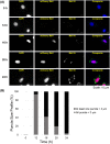
Time course of changes in viral inclusion body formation following measles virus infection. HeLa cells were infected (MOI, 5) with recombinant measles virus (MeV vac2) isogenic to the Moraten vaccine strain engineered to express mCherry protein with a nuclear localization signal. Cells were fixed with formaldehyde at the indicated time after infection, immunostained for MeV N protein or stained with DAPI for DNA, and subjected to fluorescent imaging analysis. (A) Representative images at each time point after infection. Scale bars = 10 μm. (B) Quantitation of time course analysis of inclusion body size, based on the longest path from two ends of the largest puncta in each cell. Black, at least one punctum larger than 3 μm; gray, all puncta less than 3 μm. A total of 500 cells were examined.
FIG 2
FIG 2
Measles virus N and P proteins are sufficient to mediate inclusion body formation. HeLa cells were transfected with plasmids expressing N alone, N(KRR/AAA) mutant alone, P alone, or cotransfected with N (either wild-type or mutant) and P at 1:1 mass ratio. The N(KRR/AAA) mutant possesses alanine substitutions at three residues, K180, R194, and R354. Cells were fixed 24 h after transfection, immunostained for N and P proteins as indicated or stained with DAPI for DNA, and subjected to fluorescent imaging analysis. (A) Transfection with N alone forms puncta in nucleoli in 95% of transfected cells. (B) Transfection with N(KRR/AAA) mutant alone displays a diffuse nuclear staining in all (100%) of cells. (C) Transfection with P alone exhibits a mixed pattern, with a diffuse cytoplasmic localization in 65% of cells (upper) and large irregular-shaped perinuclear puncta in 35% of cells (lower). (D) Coexpression of wild-type N and P results in spherical IB-like puncta in 95% of infected cells. (E) Coexpression of N(KRR/AAA) mutant and P results in puncta similar to panel D with wild-type N. (A to E) The % values in represent averages from the results from three independent experiments with 500 transfected cells examined in each experiment. (F and G) Representative images (F) and IB size analysis (G) at 16 h and 24 h after transfection. The analysis of IB size was based on the longest path from two ends of the largest punctum in each cell, and a total of 500 cells were examined at each time point. Scale bars = 10 μm.
FIG 3
FIG 3
Colocalization of cellular WDR5 protein to inclusion bodies containing measles virus N and P proteins in virus-infected and plasmid-transfected cells. (A and B) HeLa cells stably expressing a mCherry fusion of WDR5 were either infected (MOI, 0.2) with isogenic Moraten vac2 virus with an HA-tagged L protein (A) or cotransfected with plasmids expressing wild-type N and P MeV proteins (B). After infection or transfection for 24 h, cells were fixed, immunostained for MeV N and P protein expression or stained with DAPI for DNA, and subjected to fluorescent imaging analysis. (C) Localization of WDR5-mCherry in uninfected cells not transfected is shown as a control. Scale bars = 10 μm.
FIG 4
FIG 4
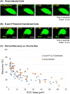
Efficient exchange occurs between inclusion body-associated and cytosolic WDR5-EGFP, as determined by FRAP. (A and B) HeLa cells stably expressing an EGFP fusion of WDR5 were either infected (MOI, 0.2) with MeV (A) or cotransfected with N and P expression constructs (B). After 24 h, the fluorescence recovery after photobleaching (FRAP) was conducted on an individual punctum by drawing a region of interest (ROI; shown as a circle), using live-cell confocal microscopy. Each ROI was photobleached, and its fluorescent recovery was monitored for 4 min. Representative images of puncta from infected cells (A) and N- and P-cotransfected cells at prephotobleaching (B); photobleached (T = 0); and postphotobleaching (T = 120 and T = 240 s). (C) An inverse correlation between the punctum size and the percent recovery of fluorescent intensity. Red circles, MeV-infected cells; blue triangles, N- and P-cotransfected cells. Note that for a given punctum size, IB-associated WDR5-EGFP displays a comparable efficiency of photobleaching recovery between MeV-infected and N- and P-cotransfected cells.
FIG 5
FIG 5
Fusion and relaxation of inclusion bodies in MeV-infected and N- and P-cotransfected cells. (A and B) HeLa cells stably expressing an EGFP fusion of WDR5 were infected (MOI, 0.2) with MeV (A) or cotransfected with N and P expression constructs (B). Live-cell imaging was performed with time-lapse microscopy to track the fusion and relaxation of IBs (designated by asterisks) using WDR5-EGFP as the marker. Snapshots at different time points show the fusion and subsequent relaxation between two IBs in MeV-infected cells (A) and N/P-cotransfected cells (B).
FIG 6
FIG 6
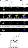
Mutational analysis identifies the C-terminal regions of measles virus N and P proteins as necessary for normal inclusion body formation. HeLa cells were cotransfected with N and P expression constructs, either wild-type or mutant, as indicated. Cells were fixed 24 h after transfection, immunostained for N and P proteins as indicated and stained with DAPI for DNA, and subjected to fluorescent imaging analysis. (A) Schematic diagram of MeV N and MeV P proteins with the amino acid positions of domains indicated. Structured and intrinsically disordered regions are indicated by boxes and solid lines, respectively. The MoRE motif located within the disordered region of N is represented by a helix. The MD and XD of P are the multimerization and X domains, respectively. (B to E) Representative images of IBs formed in cotransfected cells. The % values are averages of the results from three independent experiments, with 500 transfected cells examined in each experiment. Scale bars = 10 μm. (B) Coexpression of N (WT) and P (WT). (C) Coexpression of N (WT) and XD deletion mutant P (aa 1 to 459). (D) Coexpression of N (WT) and substitution mutant P (LFM/AAA) containing alanine substitutions within the XD domain of P (L484A, F497A, and M500A). (E) Coexpression of deletion mutant N (aa 1 to 391) and P (WT). (F) Western blot analysis of cell lysates showing expression levels of N and P proteins; 34-, 43-, 56-, and 72-kDa molecular mass markers are indicated.
FIG 7
FIG 7
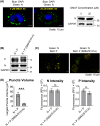
Phosphorylation of measles virus P protein modulates inclusion body size. HeLa cells were infected with MeV or cotransfected with N and P expression constructs. Cells were fixed after infection (18 h) or transfection (24 h), immunostained for N and P proteins or stained with DAPI for DNA, as indicated, and subjected to fluorescent imaging analysis. (A) Cells were infected (MOI, 0.2) and at 12 h after infection were treated with the casein kinase 2 inhibitor DMAT or the solvent DMSO vehicle for 6 h before fixation and immunostained for N protein (left). The level of N was also determined by a Western blot assay (right). (B) Western blot analysis of expression levels of wild-type N and P and the CK2 phosphorylation site double-substitution mutant, P(S86A, S151A); 34-, 56-, and 72-kDa molecular mass markers are indicated. (C) Representative confocal images of N and P cotransfection puncta formed with wild-type N plus either wild-type or mutant P(S86A, S151A). Scale bars = 10 μm. (D) The cellular expression levels of N and P proteins for cells transfected with N and P or N and mutant P(S86A, S151A) were quantified using the Imaris three-dimensional (3D) reconstruction software (represented by relative fluorescent units [RFU]). For each group, 25 cells exhibiting similar N and P levels were selected for further analysis. The volumes of the largest puncta found in each of the 25 cells were measured for both groups and averaged. The error bar represents the standard error of the mean. ***, P < 0.001.
FIG 8
FIG 8
Dynein-mediated transport promotes the formation of large inclusion bodies in measles virus-infected cells. HeLa cells at 12 h after infection (MOI, 0.2) were treated with a dynein inhibitor, either HPI-4 or dynarrestin, at the indicated concentrations for 6 h before confocal microscopy analysis. (A) The size profile of the largest punctum present in each infected cell treated with increasing concentrations of HPI-4, as indicated. Punctum size was measured as the longest path from two ends of the largest punctum in each cell. A total of 500 cells were examined under each condition. (B) Representative images of cells either untreated or treated with 80 μm HPI-4. Scale bars = 10 μm. (C) Volumes of the largest punctum found in individual cells were measured using the Imaris 3D reconstruction software. A total of 55 cells were examined under each condition. (D) Western blot analysis of lysates prepared from untreated and HPI-treated cells for N and P expression; GAPDH and α-tubulin are included for loading controls; 34-, 56-, and 72-kDa molecular mass markers are indicated. (E) Representative images of cells either untreated or treated with 30 μm dynarrestin. Scale bars = 10 μm.

References

    1. Lamb RA, Parks GD. 2013. Paramyxoviridae, p 957–995. In Knipe DM, Howley PM, Cohen JI, Griffin DE, Lamb RA, Martin MA, Racaniello VR, Roizman B (ed), Fields virology, 6th ed, vol 1 Lippincott Williams & Wilkins, Philadelphia, PA.
    1. Cattaneo R, Donohue RC, Generous AR, Navaratnarajah CK, Pfaller CK. 2019. Stronger together: multi-genome transmission of measles virus. Virus Res 265:74–79. doi: 10.1016/j.virusres.2019.03.007. - DOI - PMC - PubMed
    1. Dabbagh A, Patel MK, Dumolard L, Gacic-Dobo M, Mulders MN, Okwo-Bele J-M, Kretsinger K, Papania MJ, Rota PA, Goodson JL. 2017. Progress toward regional measles elimination—worldwide, 2000–2016. MMWR Morb Mortal Wkly Rep 66:1148. doi: 10.15585/mmwr.mm6642a6. - DOI - PMC - PubMed
    1. Griffin D. 2013. Measles, p 1042–1069. In Knipe DM, Howley PM, Cohen JI, Griffin DE, Lamb RA, Martin MA, Racaniello VR, Roizman B (ed), Fields virology, 6th ed, vol 1 Lippincott Williams & Wilkins, Philadelphia, PA.
    1. Bourhis J-M, Johansson K, Receveur-Bréchot V, Oldfield CJ, Dunker KA, Canard B, Longhi S. 2004. The C-terminal domain of measles virus nucleoprotein belongs to the class of intrinsically disordered proteins that fold upon binding to their physiological partner. Virus Res 99:157–167. doi: 10.1016/j.virusres.2003.11.007. - DOI - PubMed

Publication types