. 2019 Aug 2;9(1):11283.
doi: 10.1038/s41598-019-47445-5.

X-ray Crystallographic Structure and Oligomerization of Gloeobacter Rhodopsin

Affiliations

X-ray Crystallographic Structure and Oligomerization of Gloeobacter Rhodopsin

Takefumi Morizumi et al. Sci Rep. .

Abstract

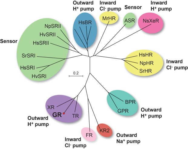
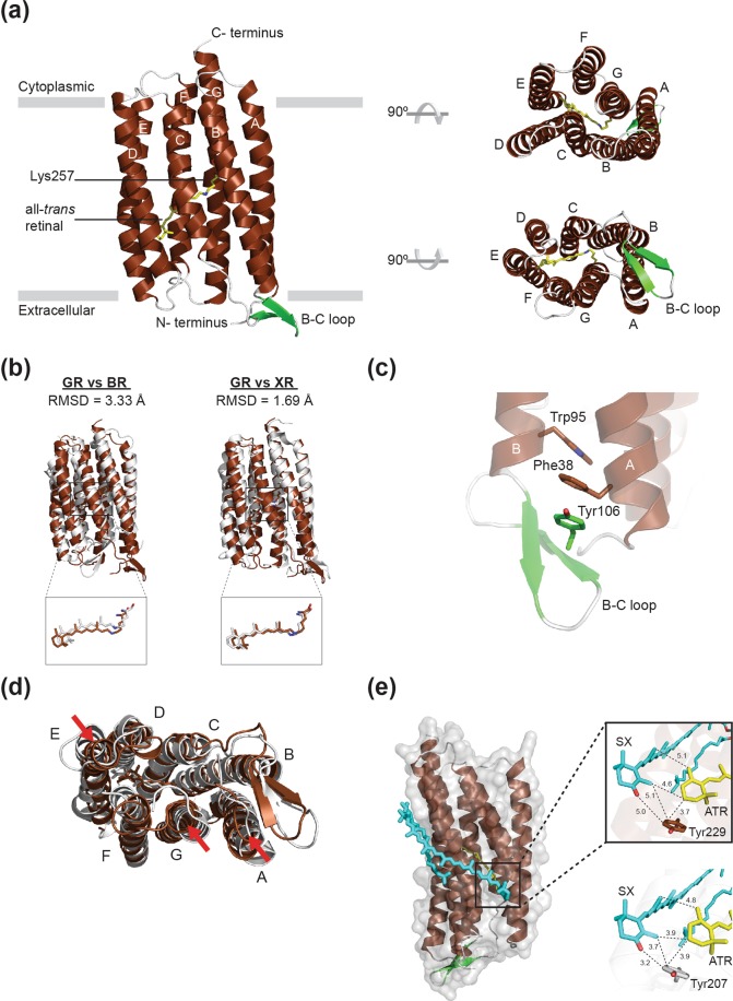
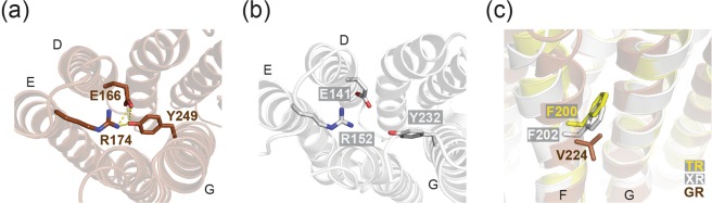
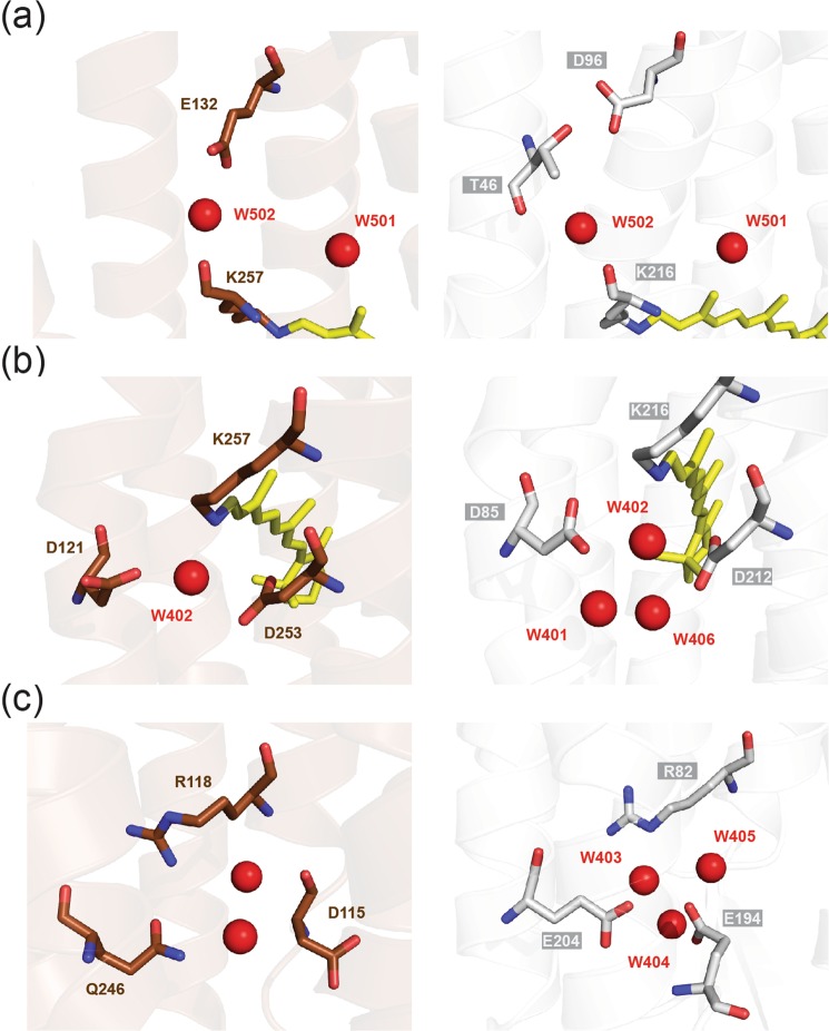
Gloeobacter rhodopsin (GR) is a cyanobacterial proton pump which can be potentially applied to optogenetics. We solved the crystal structure of GR and found that it has overall similarity to the homologous proton pump from Salinibacter ruber, xanthorhodopsin (XR). We identified distinct structural characteristics of GR's hydrogen bonding network in the transmembrane domain as well as the displacement of extracellular sides of the transmembrane helices relative to those of XR. Employing Raman spectroscopy and flash-photolysis, we found that GR in the crystals exists in a state which displays retinal conformation and photochemical cycle similar to the functional form observed in lipids. Based on the crystal structure of GR, we selected a site for spin labeling to determine GR's oligomerization state using double electron-electron resonance (DEER) spectroscopy and demonstrated the pH-dependent pentamer formation of GR. Determination of the structure of GR as well as its pentamerizing propensity enabled us to reveal the role of structural motifs (extended helices, 3-omega motif and flipped B-C loop) commonly found among light-driven bacterial pumps in oligomer formation. Here we propose a new concept to classify these pumps based on the relationship between their oligomerization propensities and these structural determinants.


Conflict of interest statement

The authors declare no competing interests.

Figures

Figure 1
Figure 1
Phylogenetic tree of microbial rhodopsins from Archaea and Eubacteria, representing the phylogenetic relationship between Gloeobacter rhodopsin and related proteins. HsBR, bacteriorhodopsin from H. salinarum; HvSRI, HsSRI, SrSRI, sensory rhodopsin I from Haloarcula vallismortis, H. salinarum, and Salinibacter ruber; HsSRII, HvSRII, NpSRII, sensory rhodopsin II from H. salinarum, Haloarcula vallismortis, Natronomonas pharaonis; MrHR, Mastigocladopsis repens halorhodopsin; ASR, Anabaena sensory rhodopsin; NsXeR, Nanosalinarum sp xenorhodopsin; HsHR, NpHR, SrHR, halorhodopsin from H. salinarum, N. pharaonis, and S. ruber; BPR, blue-absorbing proteorhodopsin; GPR, green-absorbing proteorhodopsin; KR2, sodium-pumping rhodopsin from Krokinobacter eikastus; FR, chloride-pumping rhodopsin from Fulvimarina pelagi; TR, thermophilic rhodopsin from Thermus thermophilus; GR, rhodopsin from Gloeobacter violaceus PCC 7421; XR, xanthorhodopsin from S. ruber. The evolutionary history was inferred using the Neighbor-Joining method. The optimal tree with the sum of branch length = 11.29080174 is shown. The tree is drawn to scale, with branch lengths in the same units as those of the evolutionary distances used to infer the phylogenetic tree. The evolutionary distances were computed using the Poisson correction method and are in the units of the number of amino acid substitutions per site. The analysis involved 23 amino acid sequences. All positions containing gaps and missing data were eliminated. There was a total of 189 positions in the final dataset. Evolutionary analyses were conducted with MEGA7.
Figure 2
Figure 2
Structural details of GR. (a) Overall structure of GR shown in cartoon representation viewed parallel to the membrane (left), from intracellular side (right, top) and from extracellular side (right, bottom). GR consists of seven transmembrane helices (TM A to TM G, shown in brown) connected by interhelical loops (shown in white) on both sides of the membrane. The β-strands in the B-C loop are shown in green. All-trans-retinal, depicted by stick models, is covalently linked to Lys257 via a protonated Schiff base (shown in yellow). (b) Superimposed structures of GR (brown) and H. salinarum BR (white, PDB entry 1C3W), and GR and XR (white, PDB entry 3DDL), respectively. (c) 3-omega motif of GR formed by π-stacking interactions of the side chains of aromatic residues in TM A (F38), TM B (W95) and B–C loop (Y106). (d) Extracellular side of superimposed GR (brown) and XR (white) structures with the XR-bound SX molecule (cyan). The helix displacements are indicated by red arrows. (e) Potential carotenoid binding site in GR. Salinixanthin (SX) from the crystal structure of XR is superimposed onto the structure of GR. The magnified views compare the location of SX and the retinal in GR (top) and XR (bottom).
Figure 3
Figure 3
Conserved Glu-Arg salt bridge on the extracellular side of helices D and E; E166-R174 in GR (a) and E141-R152 in XR (b). (c) Steric interference of movement of helices F and G by Phe residues on helix F in XR and TR.
Figure 4
Figure 4
Comparison of three internal cavities in proton transporting pathway of GR (brown) and BR (white). (a) Cytoplasmic side of the Schiff base region. (b) Extracellular side of the Schiff base region. (c) Cavities near the extracellular surface.
Figure 5
Figure 5
(a) UV-Vis spectra of GR at different pH conditions. Absorption maxima are indictaed. (b) Size exclusion chromatogram of GR at different pH conditions. Peak elution volumes are indictaed. (c,d) EM images of GR at pH 8.0 and 3.0, negatively stained with 2% uranyl acetate (Scale bar, 100 nm).
Figure 6
Figure 6
EPR analysis of the oligomeric states of spin labeled GR-67R1. (a) Spin-labeling site on GR viewed parallel to the membrane (left) and from the cytoplasmic side (right). The α-carbon of Gly67 is colored magenta and shown as a sphere. (bd) Superimposed structure of GR and HsBR (b) GR and KR2 (c) and BPR (d). α-carbon of the residues equivalent to Gly67 in GR are shown as a sphere. (e) Room temperature CW EPR spectra of GR-67R1 in 0.05% DDM at pH 3.0 and pH 8.0. Dipolar broadening in the pH 8.0 spectrum is clearly observed. (f) DEER distance distribution of GR-67R1 at pH 8.0. Inset: baseline-corrected DEER traces.
Figure 7
Figure 7
Summary of relationships between the evolutionally altered structural features and oligomerization propensities. (a) Phylogenetic tree and structural motifs. The important branches are numbered: extension of the interface helices as 1, acquisition of 3-omega motif and flipped B-C loop as 2 and the functional divergence in XR-type proteins as 3. (b) (upper) Crystal structures of BR (left, PDB), BPR-Med12 (middle) and GR (right) viewed parallel to the membrane. (lower) Trimeric organization of BR (left) and modelled pentameric organization of GR (right). B-C loops are colored in Red.

References

    1. Ernst OP, et al. Microbial and animal rhodopsins: structures, functions, and molecular mechanisms. Chemical reviews. 2014;114:126–163. doi: 10.1021/cr4003769. - DOI - PMC - PubMed
    1. Spudich, J. L., Yang, C. S., Jung, K. H. & Spudich, E. N. Retinylidene Proteins: Structures and Functions from Archaea to Humans. Annual review of cell and developmental biology16 (2000). - PubMed
    1. Kurihara M, Sudo Y. Microbial rhodopsins: wide distribution, rich diversity and great potential. Biophys Physicobiol. 2015;12:121–129. doi: 10.2142/biophysico.12.0_121. - DOI - PMC - PubMed
    1. Kandori H. Ion-pumping microbial rhodopsins. Front Mol Biosci. 2015;2:52. doi: 10.3389/fmolb.2015.00052. - DOI - PMC - PubMed
    1. Cao Z, et al. Novel expression and characterization of a light driven proton pump archaerhodopsin 4 in a Halobacterium salinarum strain. Biochimica et biophysica acta. 2015;1847:390–398. doi: 10.1016/j.bbabio.2014.12.008. - DOI - PubMed

Publication types

MeSH terms

Supplementary concepts