. 2019 Aug 2;9(1):11278.
doi: 10.1038/s41598-019-47643-1.

Neural indices of listening effort in noisy environments

Affiliations

Neural indices of listening effort in noisy environments

Andrew Dimitrijevic et al. Sci Rep. .

Abstract

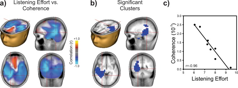
Listening in a noisy environment is challenging for individuals with normal hearing and can be a significant burden for those with hearing impairment. The extent to which this burden is alleviated by a hearing device is a major, unresolved issue for rehabilitation. Here, we found adult users of cochlear implants (CIs) self-reported listening effort during a speech-in-noise task that was positively related to alpha oscillatory activity in the left inferior frontal cortex, canonical Broca's area, and inversely related to speech envelope coherence in the 2-5 Hz range originating in the superior-temporal plane encompassing auditory cortex. Left frontal cortex coherence in the 2-5 Hz range also predicted speech-in-noise identification. These data demonstrate that neural oscillations predict both speech perception ability in noise and listening effort.


Conflict of interest statement

The authors declare no competing interests.

Figures

Figure 1
Figure 1
Attention enhances brain oscillations. (a) Grand mean time-frequency representations for the passive (left) and attentive (right) listening conditions. The digits in noise acoustic waveform is shown above each plot (light grey) indicating the timing of the stimuli relative to the time-frequency plot. The darker grey rectangle schematically shows the noise onset and offset. Oscillatory power increased (red; event-related synchronization, ERS) or decreased (blue; event-related desynchronization, ERD) as a percent change relative to baseline (−1 to 0 s). The attentive condition was characterized by greater oscillatory power change in the alpha (8–12 Hz) band, and reduced power in the beta (20–28 Hz) and gamma (35–40 Hz) bands compared to the passive listening condition. Time frequency data were averaged across all 63 electrodes. Oscillatory topography is shown on the right. (b) Beamformer source analysis over the dotted time-frequency window indicated that the alpha ERS is generated in the right posterior parietal cortex, beta ERD in left inferior frontal gyrus, and gamma ERD in bilateral anterior poles. Head and brain images were in generated in BESA Research 7.1 (http://www.besa.de/).
Figure 2
Figure 2
Listening effort is related to alpha power in left frontal regions. (a) Correlation between alpha power (for each beamformer voxel) and listening effort (NASA Task Load Index rating from 1–10) rating across all CI users. Each voxel represents a Pearson correlation coefficient. Yellow represents voxels with high positive correlations and blue represents high negative correlations. The maximum positive correlation is indicated by the cross hairs at Talairach location −39, 11, 10 corresponding to Brodman area 44, the left inferior frontal gyrus (IFG). (b) Only the left frontal and temporal clusters survived multiple comparisons corrections (p = 0.048, corrected). (c) A scatter plot between listening effort and alpha power at the left IFG voxel with correlation value of r = 0.97. Increased effort is associated with increased alpha ERS. Head and brain images were in generated in BESA Research 7.1 (http://www.besa.de/).
Figure 3
Figure 3
Listening effort is related to speech-brain coherence. (a) Correlation between listening effort and speech-brain coherence (in the 2–5 Hz range) across all CI users. Each voxel represents a Pearson correlation coefficient, as in Fig. 2. High negative correlations were observed bilaterally in the temporal lobe, including auditory cortex. The minimum correlation is indicated by the cross hairs at Talairach location -25, -45, -4 corresponding to Brodman area 37, the left fusiform gyrus. (b) Only the left temporal clusters survived multiple comparisons corrections. (p = 0.039 for cluster). (c) Relation between listening effort and speech-brain coherence power measured at the peak correlation (left fusiform gyrus voxel with correlation value of r = −0.96). Increased effort is associated with decreased speech-brain coherence. Head and brain images were in generated in BESA Research 7.1 (http://www.besa.de/).
Figure 4
Figure 4
Correct digit identification is related to DIN speech-brain coherence in left frontal regions. (a) DICS (DIN speech envelope – to whole brain at 2–5 Hz) were performed separately on correct and incorrect trials. Significant differences between correct and incorrect trials were observed in left prefrontal cortex (cross hairs at −32, 40, 16; p = 0.005). (b) Individual coherence values at voxel locations at left and right Heschl’s Gyrus (HG) and at left prefrontal cortex (cross-hairs in (a)) for correct and incorrect trials. Only the left frontal pole showed a significant difference between correct and incorrect DIN identification. Head and brain images were in generated in BESA Research 7.1 (http://www.besa.de/).

References

    1. McGarrigle R, et al. Listening effort and fatigue: what exactly are we measuring? A British Society of Audiology Cognition in Hearing Special Interest Group ‘white paper’. Int. J. Audiol. 2014;53:433–40. doi: 10.3109/14992027.2014.890296. - DOI - PubMed
    1. Pichora-Fuller MK, et al. Hearing Impairment and Cognitive Energy: The Framework for Understanding Effortful Listening (FUEL) Ear Hear. 2016;37:5S–27S. doi: 10.1097/AUD.0000000000000312. - DOI - PubMed
    1. Winn M. Rapid Release From Listening Effort Resulting From Semantic Context, and Effects of Spectral Degradation and Cochlear Implants. Trends Hear. 2016;20:1–17. - PMC - PubMed
    1. Hughes KC, Galvin KL. Measuring listening effort expended by adolescents and young adults with unilateral or bilateral cochlear implants or normal hearing. Cochlear Implants Int. 2013;14:121–129. doi: 10.1179/1754762812Y.0000000009. - DOI - PubMed
    1. Alhanbali S, Dawes P, Lloyd S, Munro KJ. Self-Reported Listening-Related Effort and Fatigue in Hearing-Impaired Adults. Ear Hear. 2016;38:e39–e48. doi: 10.1097/AUD.0000000000000361. - DOI - PubMed

Publication types