

Functional regeneration of tissue engineered skeletal muscle in vitro is dependent on the inclusion of basement membrane proteins
- PMID: 31376315
- PMCID: PMC6790946
- DOI: 10.1002/cm.21553
Functional regeneration of tissue engineered skeletal muscle in vitro is dependent on the inclusion of basement membrane proteins
Abstract
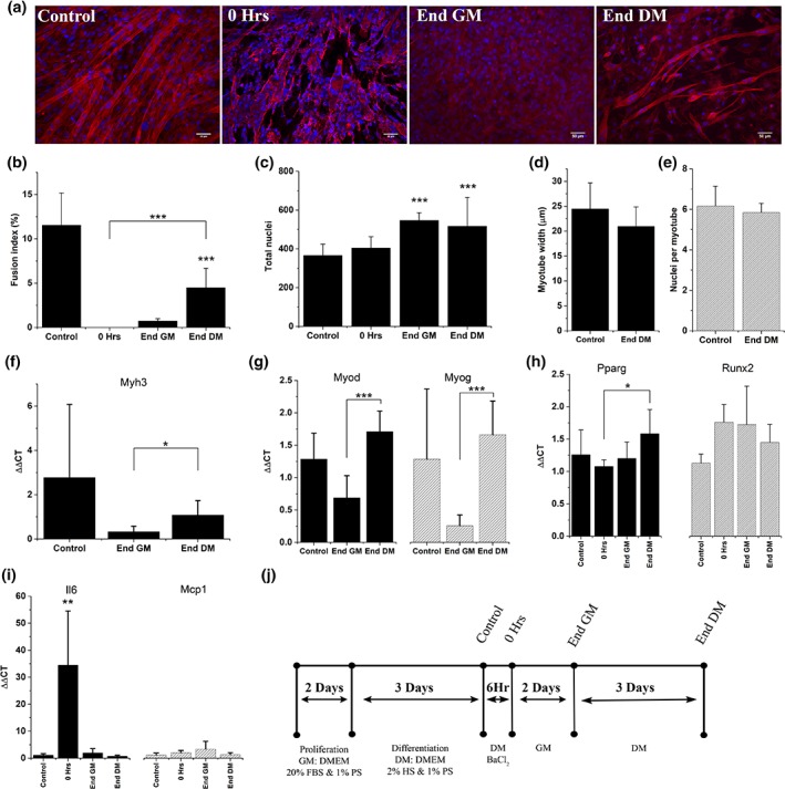
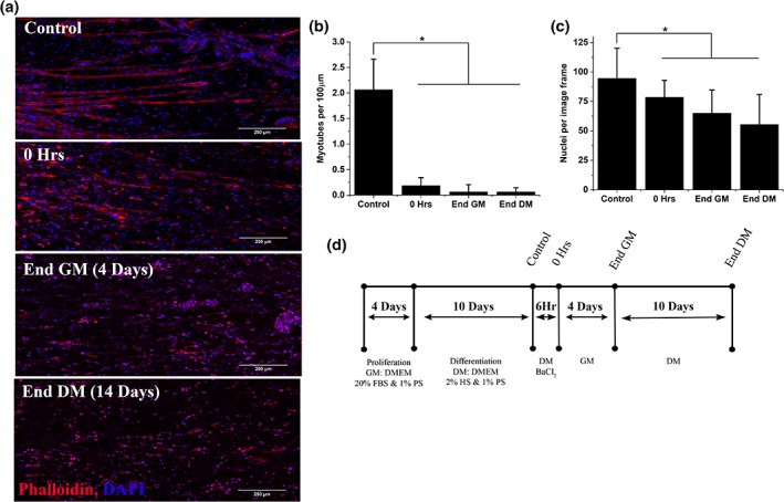
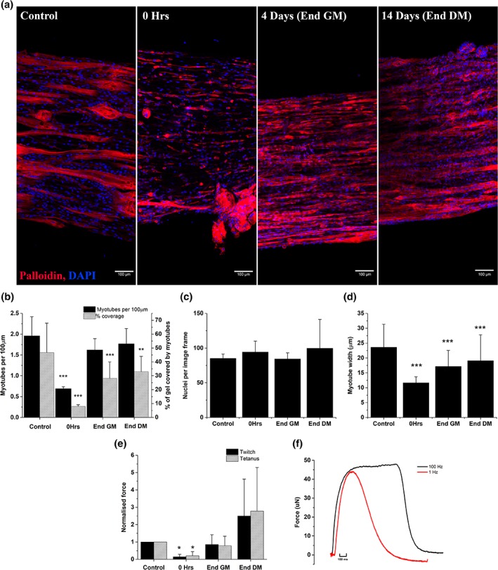
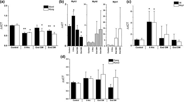
Skeletal muscle has a high regenerative capacity, injuries trigger a regenerative program which restores tissue function to a level indistinguishable to the pre-injury state. However, in some cases where significant trauma occurs, such as injuries seen in military populations, the regenerative process is overwhelmed and cannot restore full function. Limited clinical interventions exist which can be used to promote regeneration and prevent the formation of non-regenerative defects following severe skeletal muscle trauma. Robust and reproducible techniques for modelling complex tissue responses are essential to promote the discovery of effective clinical interventions. Tissue engineering has been highlighted as an alternative method, allowing the generation of three-dimensional in vivo like tissues without laboratory animals. Reducing the requirement for animal models promotes rapid screening of potential clinical interventions, as these models are more easily manipulated, genetically and pharmacologically, and reduce the associated cost and complexity, whilst increasing access to models for laboratories without animal facilities. In this study, an in vitro chemical injury using barium chloride is validated using the C2C12 myoblast cell line, and is shown to selectively remove multinucleated myotubes, whilst retaining a regenerative mononuclear cell population. Monolayer cultures showed limited regenerative capacity, with basement membrane supplementation or extended regenerative time incapable of improving the regenerative response. Conversely tissue engineered skeletal muscles, supplemented with basement membrane proteins, showed full functional regeneration, and a broader in vivo like inflammatory response. This work outlines a freely available and open access methodology to produce a cell line-based tissue engineered model of skeletal muscle regeneration.
Keywords: bioengineering; regeneration; skeletal muscle physiology; tissue engineering.
© 2019 The Authors. Cytoskeleton published by Wiley Periodicals, Inc.
Conflict of interest statement
The authors have no conflict of interest to declare.
Figures
Similar articles
-
Bioengineered human skeletal muscle capable of functional regeneration.BMC Biol. 2020 Oct 20;18(1):145. doi: 10.1186/s12915-020-00884-3. BMC Biol. 2020. PMID: 33081771 Free PMC article.
-
Growth factor supplemented matrigel improves ectopic skeletal muscle formation--a cell therapy approach.J Cell Physiol. 2001 Feb;186(2):183-92. doi: 10.1002/1097-4652(200102)186:2<183::AID-JCP1020>3.0.CO;2-Q. J Cell Physiol. 2001. PMID: 11169455
-
Effects of type IV collagen on myogenic characteristics of IGF-I gene-engineered myoblasts.J Biosci Bioeng. 2015 May;119(5):596-603. doi: 10.1016/j.jbiosc.2014.10.008. Epub 2014 Nov 21. J Biosci Bioeng. 2015. PMID: 25454061
-
Ionic medicine: Exploiting metallic ions to stimulate skeletal muscle tissue regeneration.Acta Biomater. 2024 Dec;190:1-23. doi: 10.1016/j.actbio.2024.10.033. Epub 2024 Oct 23. Acta Biomater. 2024. PMID: 39454933 Review.
-
Galectin-1 is a novel factor that regulates myotube growth in regenerating skeletal muscles.Curr Drug Targets. 2005 Jun;6(4):395-405. doi: 10.2174/1389450054021918. Curr Drug Targets. 2005. PMID: 16026258 Review.
Cited by
-
Spatiotemporal single-cell tracking analysis in 3D tissues to reveal heterogeneous cellular response to mechanical stimuli.Sci Adv. 2023 Oct 13;9(41):eadf9917. doi: 10.1126/sciadv.adf9917. Epub 2023 Oct 13. Sci Adv. 2023. PMID: 37831766 Free PMC article.
-
A concise in vitro model for evaluating interactions between macrophage and skeletal muscle cells during muscle regeneration.Front Cell Dev Biol. 2023 May 18;11:1022081. doi: 10.3389/fcell.2023.1022081. eCollection 2023. Front Cell Dev Biol. 2023. PMID: 37274738 Free PMC article.
-
Ginkgolide B facilitates muscle regeneration via rejuvenating osteocalcin-mediated bone-to-muscle modulation in aged mice.J Cachexia Sarcopenia Muscle. 2023 Jun;14(3):1349-1364. doi: 10.1002/jcsm.13228. Epub 2023 Apr 19. J Cachexia Sarcopenia Muscle. 2023. PMID: 37076950 Free PMC article.
-
Bioengineered human skeletal muscle capable of functional regeneration.BMC Biol. 2020 Oct 20;18(1):145. doi: 10.1186/s12915-020-00884-3. BMC Biol. 2020. PMID: 33081771 Free PMC article.
-
Engineered M13 phage as a novel therapeutic bionanomaterial for clinical applications: From tissue regeneration to cancer therapy.Mater Today Bio. 2023 Mar 24;20:100612. doi: 10.1016/j.mtbio.2023.100612. eCollection 2023 Jun. Mater Today Bio. 2023. PMID: 37063776 Free PMC article. Review.
References
-
- Arnold, L. , Henry, A. , Poron, F. , Baba‐Amer, Y. , van Rooijen, N. , Plonquet, A. , … Chazaud, B. (2007). Inflammatory monocytes recruited after skeletal muscle injury switch into antiinflammatory macrophages to support myogenesis. The Journal of Experimental Medicine, 204(5), 1057–1069. 10.1084/jem.20070075 - DOI - PMC - PubMed
Publication types
MeSH terms
Substances
Grants and funding
LinkOut - more resources
Full Text Sources
