

Incorporation of Silver Sulfadiazine into An Electrospun Composite of Polycaprolactone as An Antibacterial Scaffold for Wound Healing in Rats
- PMID: 31376319
- PMCID: PMC6722444
- DOI: 10.22074/cellj.2020.6341
Incorporation of Silver Sulfadiazine into An Electrospun Composite of Polycaprolactone as An Antibacterial Scaffold for Wound Healing in Rats
Abstract
Objective: Fabrication of an antibiotic-loaded scaffold with controlled release properties for wound dressing is one of tissue engineering challenges. The aim of this study was to evaluate the wound-healing effectiveness of 500-μm thick polycaprolactone (PCL) nanofibrous mat containing silver sulfadiazine (SSD) as an antibacterial agent.
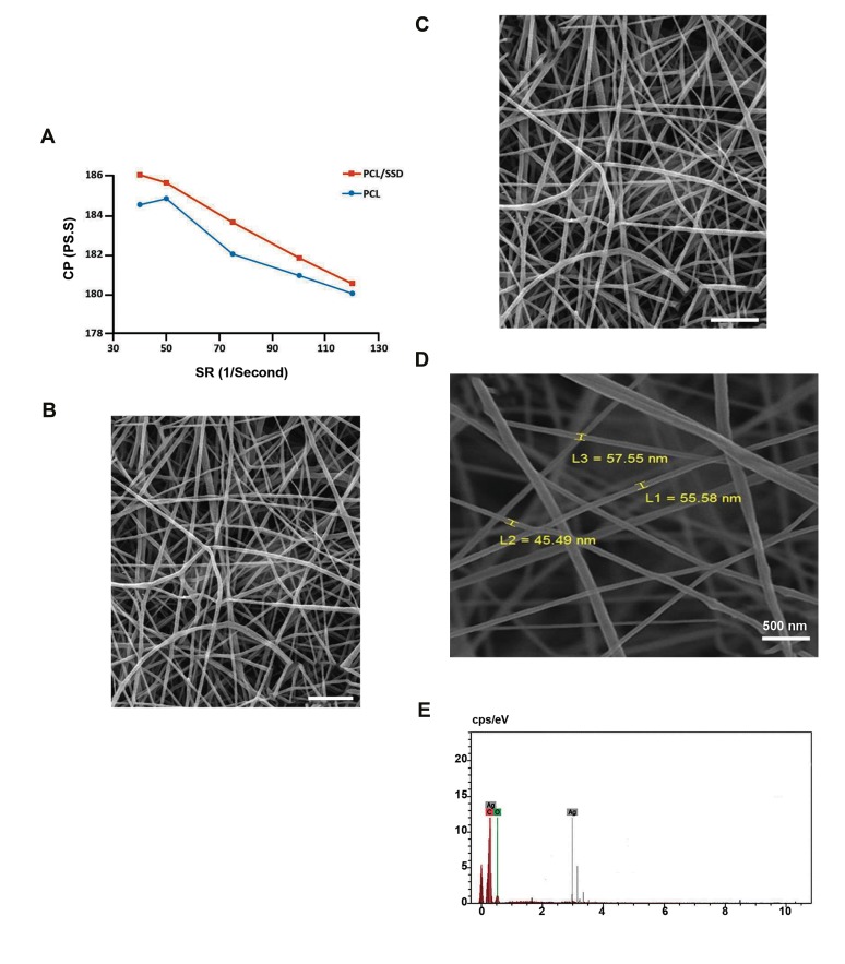
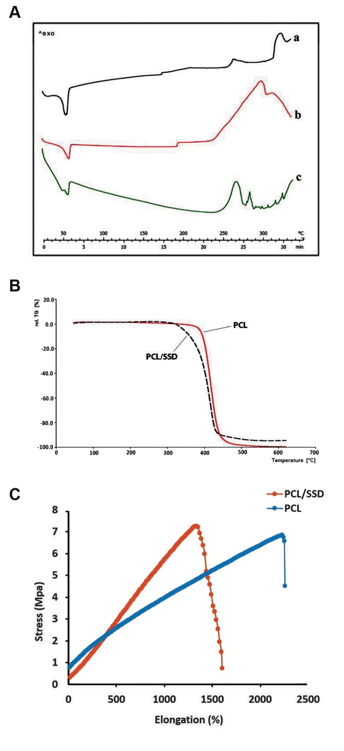
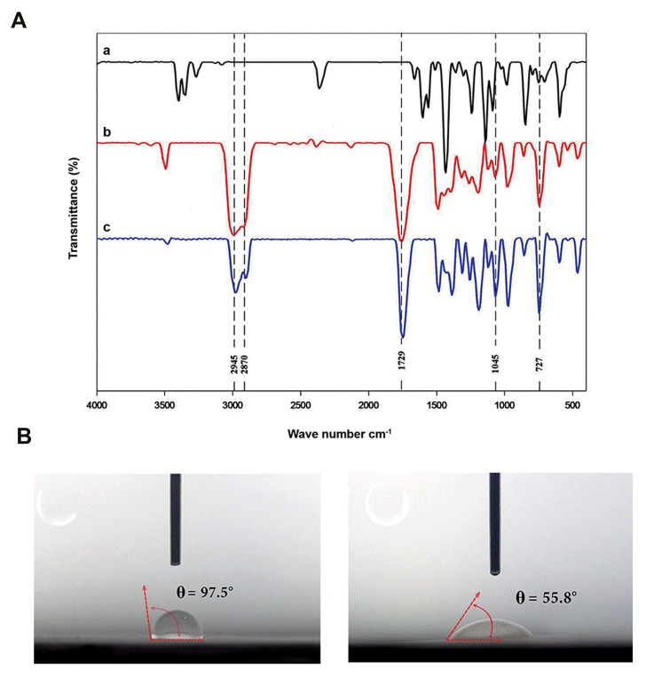
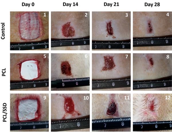
Materials and methods: In this experimental study, an electrospun membrane of PCL nanofibrous mat containing 0.3% weight SSD with 500 μm thickness, was prepared. Morphological and thermomechanical characteristics of nanofibers were evaluated. Drug content and drug release properties as well as the surface hydrophobicity of the nanofibrous membrane were determined. Antimicrobial properties and cellular viability of the scaffold were also examined. A full thickness wound of 400 mm2 was created in rats, to evaluate the wound-healing effects of PCL/SSD blend in comparison with PCL and vaseline gas used as the control group.
Results: SSD at a concentration of 0.3% improved physicochemical properties of PCL. This concentration of SSD did not inhibit the attachment of human dermal fibroblasts (HDFs) to nanofibers in vitro, but showed antibacterial activity against Gram-positive Staphylococcus aureus (ST) and Gram-negative Pseudomonas aeruginosa (PS). Overall, results showed that SSD improves characteristics of PCL nanofibrous film and improves wound-healing process in one-week earlier compared to control.
Conclusion: Cytotoxicity of SSD in fabricated nanofibrous mat is a critical challenge in designing an effective wound dressing that neutralizes cellular toxicity and improves antimicrobial activity. The PCL/SSD nanofibrous membrane with 500- μm thickness and 0.3% (w/v) SSD showed applicable characteristics as a wound dressing and it accelerated wound healing process in vivo.
Keywords: Nanofibers; Polycaprolactone; Silver Sulfadiazine; Tissue Engineering; Wound Healing.
Copyright© by Royan Institute. All rights reserved.
Conflict of interest statement
There is no conflict of interest in this study.
Figures

Similar articles
-
Application of polycaprolactone, chitosan, and collagen composite as a nanofibrous mat loaded with silver sulfadiazine and growth factors for wound dressing.Artif Organs. 2019 Apr;43(4):413-423. doi: 10.1111/aor.13369. Epub 2018 Nov 22. Artif Organs. 2019. PMID: 30311249
-
Antimicrobial Wound Dressing Containing Silver Sulfadiazine With High Biocompatibility: In Vitro Study.Artif Organs. 2016 Aug;40(8):765-73. doi: 10.1111/aor.12682. Epub 2016 Apr 20. Artif Organs. 2016. PMID: 27094090
-
Harnessing biocompatible nanofibers and silver nanoparticles for wound healing: Sandwich wound dressing versus commercial silver sulfadiazine dressing.Mater Sci Eng C Mater Biol Appl. 2021 Sep;128:112342. doi: 10.1016/j.msec.2021.112342. Epub 2021 Jul 30. Mater Sci Eng C Mater Biol Appl. 2021. PMID: 34474892
-
Electrospun Nanofibers/Nanofibrous Scaffolds Loaded with Silver Nanoparticles as Effective Antibacterial Wound Dressing Materials.Pharmaceutics. 2021 Jun 26;13(7):964. doi: 10.3390/pharmaceutics13070964. Pharmaceutics. 2021. PMID: 34206857 Free PMC article. Review.
-
Electrospun Polycaprolactone Nanofibers: Current Research and Applications in Biomedical Application.Adv Pharm Bull. 2022 Aug;12(4):658-672. doi: 10.34172/apb.2022.070. Epub 2021 Oct 3. Adv Pharm Bull. 2022. PMID: 36415646 Free PMC article. Review.
Cited by
-
Surface Characterization and Physiochemical Evaluation of P(3HB-co-4HB)-Collagen Peptide Scaffolds with Silver Sulfadiazine as Antimicrobial Agent for Potential Infection-Resistance Biomaterial.Polymers (Basel). 2021 Jul 26;13(15):2454. doi: 10.3390/polym13152454. Polymers (Basel). 2021. PMID: 34372060 Free PMC article.
-
Antibacterial and antioxidant double-layered nanofibrous mat promotes wound healing in diabetic rats.Sci Rep. 2023 Feb 23;13(1):3166. doi: 10.1038/s41598-023-30240-8. Sci Rep. 2023. PMID: 36823173 Free PMC article.
-
Fabrication and Characterization of Glycerol/Chitosan/Polyvinyl Alcohol-Based Transparent Hydrogel Films Loaded with Silver Nanoparticles for Antibacterial Wound Dressing Applications.Adv Biomed Res. 2021 Jan 27;10:4. doi: 10.4103/abr.abr_211_20. eCollection 2021. Adv Biomed Res. 2021. PMID: 33959561 Free PMC article.
-
Polycaprolactone/gelatin electrospun nanofibres containing biologically produced tellurium nanoparticles as a potential wound dressing scaffold: Physicochemical, mechanical, and biological characterisation.IET Nanobiotechnol. 2021 May;15(3):277-290. doi: 10.1049/nbt2.12020. Epub 2021 Feb 7. IET Nanobiotechnol. 2021. PMID: 34694673 Free PMC article.
-
Antimicrobial stewardship of antiseptics that are pertinent to wounds: the need for a united approach.JAC Antimicrob Resist. 2021 Mar 25;3(1):dlab027. doi: 10.1093/jacamr/dlab027. eCollection 2021 Mar. JAC Antimicrob Resist. 2021. PMID: 34223101 Free PMC article. Review.
References
-
- Klama-Baryła A, Kitala D, Łabuś W, Kraut M, Glik J, Nowak M, et al. Autologous and allogeneic skin cell grafts in the treatment of severely burned patients: retrospective clinical study. Transplant Proc. 2018;50(7):2179–2187. - PubMed
-
- Mahoney C, McCullough MB, Sankar J, Bhattarai N. Nanofibrous structure of chitosan for biomedical applications. J Nanomedic Biotherapeu Discover. 2012;2(1):102–102.
-
- Leung V, Ko F. Biomedical applications of nanofibers. Polym Adv Technol. 2011;22(3):350–365.
-
- Edwards A, Jarvis D, Hopkins T, Pixley S, Bhattarai N. Poly (ε‐ caprolactone)/keratin‐based composite nanofibers for biomedical applications. J Biomed Mater Res B Appl Biomater. 2015;103(1):21–30. - PubMed
LinkOut - more resources
Full Text Sources