. 2019 Aug 3:25:5776-5784.
doi: 10.12659/MSM.912106.

The Neuroprotective Effect of Steroid Receptor Coactivator-Interacting Protein (SIP) in Astrocyte Model of 1-Methyl-4-Phenylpyridinium (MPP⁺)-Induced Parkinson's Disease

Affiliations

The Neuroprotective Effect of Steroid Receptor Coactivator-Interacting Protein (SIP) in Astrocyte Model of 1-Methyl-4-Phenylpyridinium (MPP⁺)-Induced Parkinson's Disease

Mingwei Qu. Med Sci Monit. .

Abstract

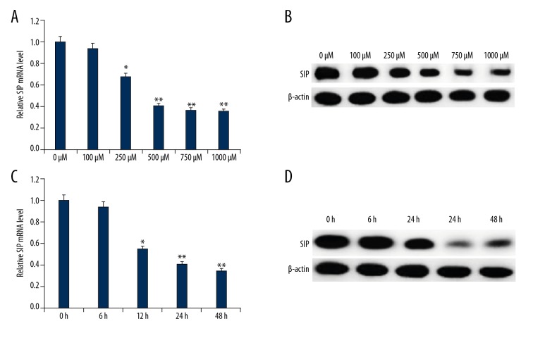
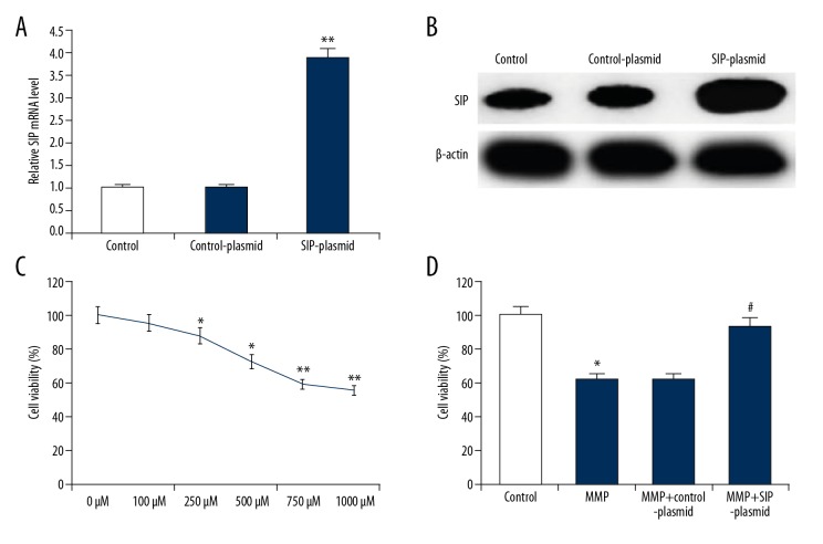
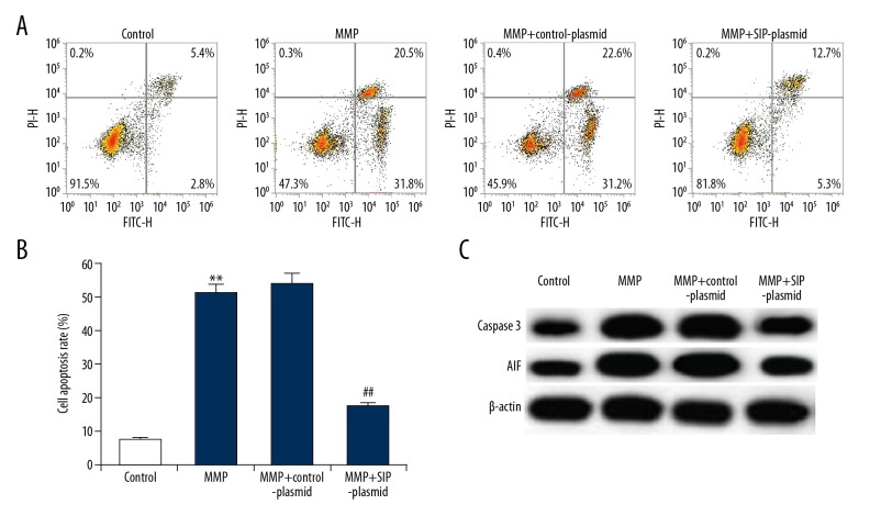
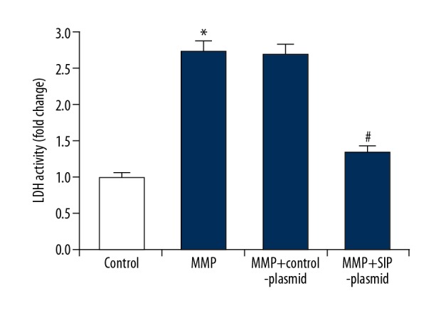
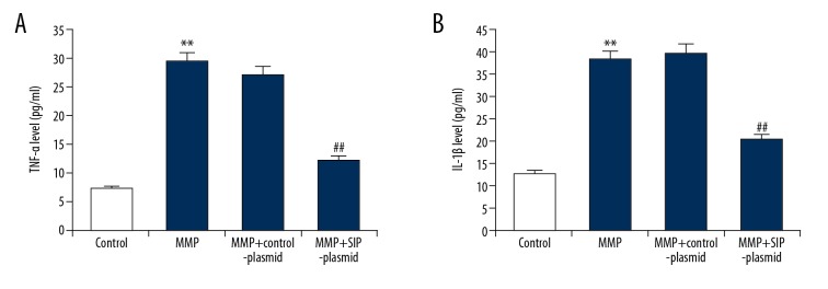
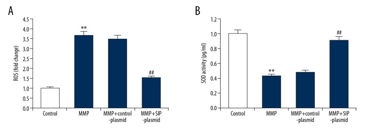
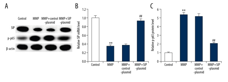
BACKGROUND The purpose of this study was to investigate the role and mechanism of steroid receptor coactivator-interacting protein (SIP) in an astrocyte model of 1-methyl-4-phenylpyridinium (MPP⁺)-induced Parkinson's disease. MATERIAL AND METHODS To perform our study, a Parkinson's disease cell model was established by treating the rat glioblastoma cell line C6 with MPP⁺. SIP was overexpressed in C6 cells using SIP-plasmid. Cell viability and apoptosis were analyzed using MTT assay and flow cytometer respectively. Tumor necrosis factor (TNF)-alpha and interleukin (IL)-1ß levels were detected using enzyme linked immunosorbent assay and quantitative reverse transcription PCR. Besides, lactate dehydrogenase (LDH) release, reactive oxygen species (ROS) production, and superoxide dismutase (SOD) enzyme activity were determined in the present study. For protein and mRNA detection, western blot assay, and qRT-PCR were performed respectively. RESULTS SIP was decreased in MPP⁺-induced C6 cells. SIP overexpression relieved MPP⁺-induced cytotoxicity of C6 cells, displayed as increased cell viability and reduced cell apoptosis and reduced LDH release. Besides, SIP inhibited MPP⁺-induced inflammatory response and oxidative stress, evidenced by decreased levels of inflammatory factors (TNF-alpha and IL-1ß), reduced ROS generation and enhanced SOD activity. Moreover, MPP⁺-induced nuclear factor-kappaB activation was inhibited by SIP overexpression. CONCLUSIONS SIP was downregulated in Parkinson's disease and it played a protective role in the development Parkinson's disease, thus may be a promising target for Parkinson's disease treatment.


Conflict of interest statement

Conflict of interests

None.

Figures

Figure 1
Figure 1
SIP expression in MMP+ treated C6 cells. C6 cells were treated with various concentrations of MPP+ (0, 100, 250, 500, 750, and 1000 μM) for 24 hours, then SIP mRNA (A) and protein (B) expression level in C6 cells was detected using qRT-PCR and western blotting, respectively. C6 cells were treated with 500 μM MPP+ for indicated times (0, 6, 12, 24, and 48 hours), then SIP mRNA (C) and protein (D) expression level in C6 cells was detected using qRT-PCR and western blotting, respectively. Data were displayed as mean ±SD. *, ** P<0.05, 0.01 versus control. SIP – steroid receptor coactivator-interacting protein; qRT-PCR – quantitative reverse transcription polymerase chain reaction; MPP+ – 1-methyl-4-phenylpyridinium; SD – standard deviation.
Figure 2
Figure 2
Effect of SIP on MMP+ treated C6 cell viability. (A) C6 cells were transfected with control-plasmid or SIP-plasmid for 24 hours, then mRNA level of SIP was measure by qRT-PCR. (B) C6 cells were transfected with control-plasmid or SIP-plasmid for 24 hours, then protein levels of SIP were measure by western blotting. (C) C6 cells were treated with various concentrations of MPP+ (0, 100, 250, 500, 750, and 1000 μM) for 24 hours, then cell viability was detected using MTT assay. (D) C6 cells were transfected with control-plasmid and SIP-plasmid for 2 hours prior to treatment with 500 μM MPP+ for 24 hours, then cell viability was detected using MTT assay. Data were displayed as mean ±SD. *, ** P<0.05, 0.01 versus control. # P<0.05 versus MMP+ treatment alone group. SIP – steroid receptor coactivator-interacting protein; qRT-PCR – quantitative reverse transcription polymerase chain reaction; MPP+ – 1-methyl-4-phenylpyridinium; SD – standard deviation.
Figure 3
Figure 3
Effect of SIP on MMP+ treated C6 cell apoptosis. C6 cells were transfected with control-plasmid and SIP-plasmid for 2 hours prior to treatment with 500 μM MPP+ for 24 hours, then cell apoptosis was analyzed by FCM (A); and the cell apoptosis rate was calculated and presented (B). The protein level of caspase-3 and AIF was detected using western blotting (C). Data were displayed as mean ±SD. ** P<0.01 versus control. ## P<0.01 versus MMP+ treatment alone group. SIP – steroid receptor coactivator-interacting protein; MPP+ – 1-methyl-4-phenylpyridinium; FCM – flow cytometer; AIF – apoptosis-inducing factor; SD – standard deviation.
Figure 4
Figure 4
Effect of SIP on LDH activity in MMP+ treated C6 cells. C6 cells were transfected with control-plasmid and SIP-plasmid for 2 hours prior to treatment with 500 μM MPP+ for 24 hours, then LDH was measured and the data were presented as fold of the control group. Data were displayed as mean ±SD. * P<0.05 versus control. # P<0.05 versus MMP+ treatment alone group. SIP – steroid receptor coactivator-interacting protein; MPP+ – 1-methyl-4-phenylpyridinium; LDH – lactate dehydrogenase; SD – standard deviation.
Figure 5
Figure 5
(A, B) Effect of SIP on TNF-α and IL-1β production in MMP+ treated C6 cells. C6 cells were transfected with control-plasmid and SIP-plasmid for 2 hours prior to treatment with 500 μM MPP+ for 24 hours, then the level of TNF-α and IL-1β in the supernatants of C6 cells was detected using ELISA. Data were displayed as mean ±SD. ** P<0.01 versus control. ## P<0.01 versus MMP+ treatment alone group. SIP – steroid receptor coactivator-interacting protein; TNF – tumor necrosis factor; IL – interleukin; MPP+ – 1-methyl-4-phenylpyridinium; ELISA – enzyme linked immunosorbent assay; SD – standard deviation.
Figure 6
Figure 6
(A, B) Effect of SIP on oxidative stress in MMP+ treated C6 cells. C6 cells were transfected with control-plasmid and SIP-plasmid for 2 hours prior to treatment with 500 μM MPP+ for 24 hours, then ROS generation and antioxidant enzymes SOD activity were determined respectively. Data were presented as fold of the control group. Data were displayed as mean ±SD. ** P<0.01 versus control. ## P<0.01 versus MMP+ treatment alone group. SIP – steroid receptor coactivator-interacting protein; MPP+ – 1-methyl-4-phenylpyridinium; ROS – reactive oxygen species; SOD – superoxide dismutase; SD – standard deviation.
Figure 7
Figure 7
Effect of SIP on NF-κB pathway in MMP+ treated C6 cells. C6 cells were transfected with control-plasmid and SIP-plasmid for 2 hours prior to treatment with 500 μM MPP+ for 24 hours, then SIP protein (A) and mRNA (B) level was detected using qRT-PCR and western blotting, and p-p65 protein (A, C) level was measured using western blot assay. Data were displayed as mean ±SD. ** P<0.01 versus control. ## P<0.01 versus MMP+ treatment alone group. SIP – steroid receptor coactivator-interacting protein; NF-κB – nuclear factor-κB; MPP+ – 1-methyl-4-phenylpyridinium; qRT-PCR – quantitative reverse transcription polymerase chain reaction; SD – standard deviation.

Similar articles

Cited by

References

    1. Valera E, Masliah E. Therapeutic approaches in Parkinson’s disease and related disorders. J Neurochem. 2016;139:346–52. - PMC - PubMed
    1. Wang Q, Liu Y, Zhou J. Neuroinflammation in Parkinson’s disease and its potential as therapeutic target. Transl Neurodegener. 2015;4:19. - PMC - PubMed
    1. He Y. Imaging brain networks in neurodegenerative diseases. CNS Neurosci Ther. 2015;21:751–53. - PMC - PubMed
    1. Baggio HC, Segura B, Junque C. Resting-state functional brain networks in Parkinson’s disease. CNS Neurosci Ther. 2015;21:793–801. - PMC - PubMed
    1. Katunina EA, Titova NV, Malykhina EA, et al. [Oxidative stress and Parkinson’s disease: Mechanisms and perspectives of treatment]. Zh Nevrol Psikhiatr Im S S Korsakova. 2015;115:141–45. [in Russian] - PubMed

MeSH terms

Substances