

A cell line resistant to avian leukosis virus subgroup B infection
- PMID: 31376350
- PMCID: PMC6771772
- DOI: 10.3382/ps/pez414
A cell line resistant to avian leukosis virus subgroup B infection
Abstract
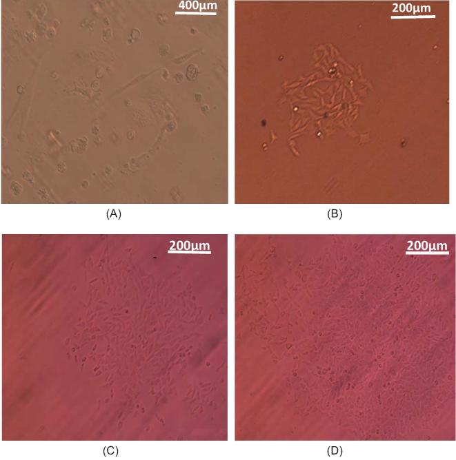
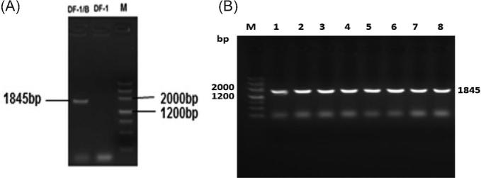
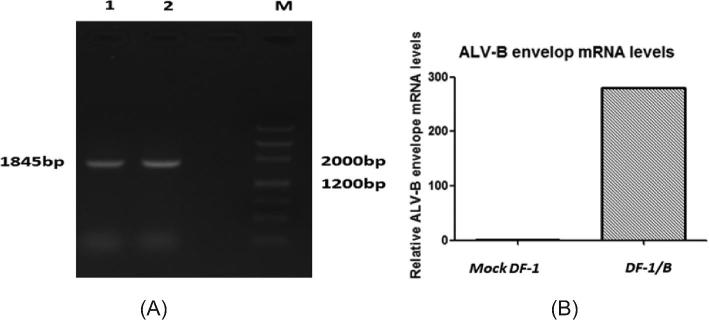
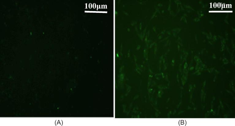
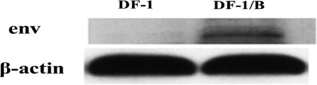
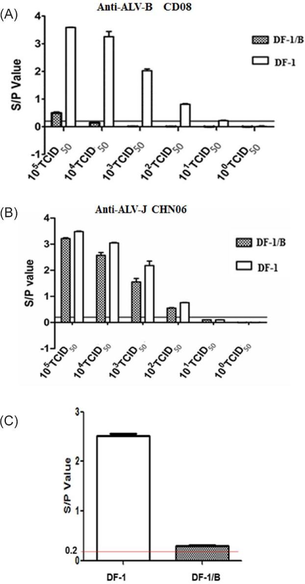
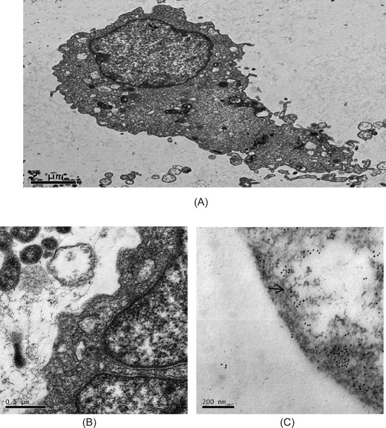
The expression of env proteins that bind to viral cell receptors on avian leukosis virus (ALV)-susceptible cells can block ALV infection. In this study, we constructed a cell line (DF-1/B) by expressing the ALV-B env protein in DF-1 cells. PCR, immune fluorescence assay, Western blot, and immune electron microscopy results showed that the env gene can be stably expressed in DF-1cells and the env protein could be detected on the DF-1 cell membrane. An antiviral experiment concluded that the DF-1/B cell line could be resistant to 1 × 10⁴ TCID50 ALV-B virus infection, but had no inhibitory effect on other subgroup ALV. This means that the DF-1/B cell line is specifically resistant to ALV-B and can be used as a tool for ALV-B diagnosis.
Keywords: env protein; ALV-B; cell line.
© The Author(s) 2019. Published by Oxford University Press on behalf of Poultry Science Association.
Figures
Similar articles
-
The construction and application of a cell line resistant to novel subgroup avian leukosis virus (ALV-K) infection.Arch Virol. 2018 Jan;163(1):89-98. doi: 10.1007/s00705-017-3563-2. Epub 2017 Oct 6. Arch Virol. 2018. PMID: 28986681 Free PMC article.
-
Inhibition of avian leukosis virus subgroup J replication by miRNA targeted against env.Virus Genes. 2013 Aug;47(1):34-41. doi: 10.1007/s11262-013-0906-2. Epub 2013 Apr 2. Virus Genes. 2013. PMID: 23546824 Free PMC article.
-
The Bipartite Sequence Motif in the N and C Termini of gp85 of Subgroup J Avian Leukosis Virus Plays a Crucial Role in Receptor Binding and Viral Entry.J Virol. 2020 Oct 27;94(22):e01232-20. doi: 10.1128/JVI.01232-20. Print 2020 Oct 27. J Virol. 2020. PMID: 32878894 Free PMC article.
-
Current knowledge on the epidemiology and prevention of Avian leukosis virus in China.Poult Sci. 2024 Sep;103(9):104009. doi: 10.1016/j.psj.2024.104009. Epub 2024 Jun 25. Poult Sci. 2024. PMID: 39002365 Free PMC article. Review.
-
Advances on genetic and genomic studies of ALV resistance.J Anim Sci Biotechnol. 2022 Oct 11;13(1):123. doi: 10.1186/s40104-022-00769-1. J Anim Sci Biotechnol. 2022. PMID: 36217167 Free PMC article. Review.
Cited by
-
Unraveling the Nrf2-ARE Signaling Pathway in the DF-1 Chicken Fibroblast Cell Line: Insights into T-2 Toxin-Induced Oxidative Stress Regulation.Toxins (Basel). 2023 Oct 25;15(11):627. doi: 10.3390/toxins15110627. Toxins (Basel). 2023. PMID: 37999490 Free PMC article.
References
-
- Cui N., Su S., Chen Z., Zhao X., Cui Z.. 2014. Genomic sequence analysis and biological characteristics of a rescued clone of avian leukosis virus strain JS11C1, isolated from indigenous chickens. J. Gen. Virol. 95:2512. - PubMed
-
- Hang B. L., Mei M., Fan Z. J.. 2013. Poultry Leukosis [M]. China Agricultural Science and Technology Press, Beijing.
-
- He C., Meng F. F., Chang S., Zhao P., Cui Z. Z.. 2013. Antigen ELISA Kit and Colloidal Gold Test Strips for the Detection of Avian Leukosis Virus Comparative Study [D]. Shandong Agricultural University; (in Chinese).
-
- Holmen S. L., Federspiel M. J.. 2000. Selection of a subgroup A avian leukosis virus [ALV(A)] envelope resistant to soluble ALV(A) surface glycoprotein. Virology 273:364–373. - PubMed
MeSH terms
Substances
LinkOut - more resources
Full Text Sources
Research Materials
Miscellaneous
