

Acute esophageal necrosis: Case report of an unknown entity
- PMID: 31376741
- PMCID: PMC6677686
- DOI: 10.1016/j.ijscr.2019.07.041
Acute esophageal necrosis: Case report of an unknown entity
Abstract
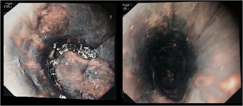
Introduction: Acute Esophageal Necrosis Syndrome (AENS) is a rare and unknown clinical entity, defined as a diffuse circumferential black-appearing friable esophageal mucosa going from the distal esophageal mucosa to the gastroesophageal (GE) junction. Esophagogastroduodenoscopy (EGD) remains the gold standard in making diagnosis.
Presentation of case: We report here the case of a 45-year-old man with necrosis of the esophagus treated conservatively. Regression of the lesion but persistence of ulcerations were seen on the endoscopic follow-up. Distal esophageal stenosis was then diagnosed and treated by endoscopic dilation.
Discussion: Diagnosis of AENS must be considered when an old patient, with multiple comorbidities, presents an upper digestive hemorrhage. Upper endoscopy is mandatory. Treatment is in most of the cases conservative. Esophageal stenosis is a frequent complication.
Conclusion: Although AENS is a rare clinical entity, it should not be dismissed by doctors, avoiding useless surgical management. This pathology remains nevertheless associated with a considerable mortality rate.
Keywords: Acute esophageal necrosis; Black esophagus; Endoscopy; Esophagus.
Copyright © 2019. Published by Elsevier Ltd.
Conflict of interest statement
None.
Figures
References
-
- Goldenberg S.P., Wain S.L., Marignani P. Acute necrotizing esophagitis. Gastroenterology. 1990;98(February (2)):493–496. - PubMed
-
- Lacy B.E., Toor A., Bensen S.P., Rothstein R.I., Maheshwari Y. Acute esophageal necrosis: report of two cases and a review of the literature. Gastrointest. Endosc. 1999;49(April (4 Pt. 1)):527–532. - PubMed
-
- Ben Soussan E., Savoye G., Hochain P., Hervé S., Antonietti M., Lemoine F., Ducrotté P. Acute esophageal necrosis: a 1-year prospective study. Gastrointest. Endosc. 2002;56(August (2)):213–217. - PubMed
-
- Moretó M., Ojembarrena E., Zaballa M., Tánago J.G., Ibánez S. Idiopathic acute esophageal necrosis: not necessarily a terminal event. Endoscopy. 1993;25(October (8)):534–538. - PubMed
LinkOut - more resources
Full Text Sources
Research Materials
