. 2019 Aug 22;75(4):835-848.e8.
doi: 10.1016/j.molcel.2019.06.031. Epub 2019 Aug 1.

MISTERMINATE Mechanistically Links Mitochondrial Dysfunction with Proteostasis Failure

Affiliations

MISTERMINATE Mechanistically Links Mitochondrial Dysfunction with Proteostasis Failure

Zhihao Wu et al. Mol Cell. .

Abstract

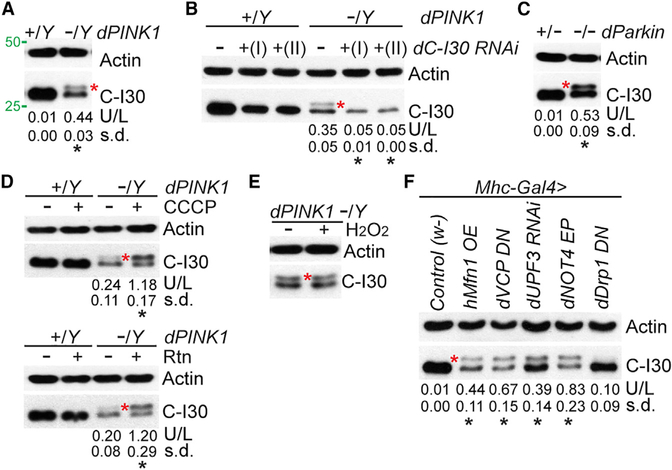
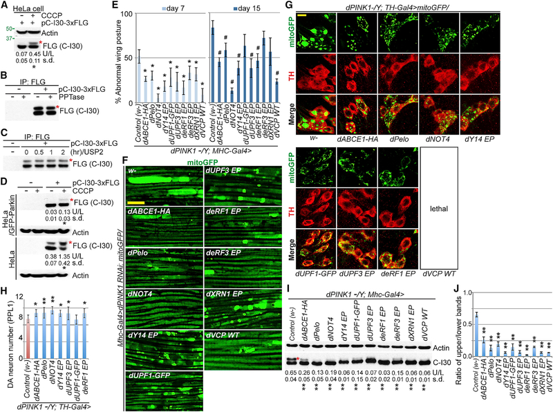
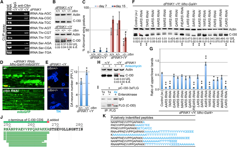
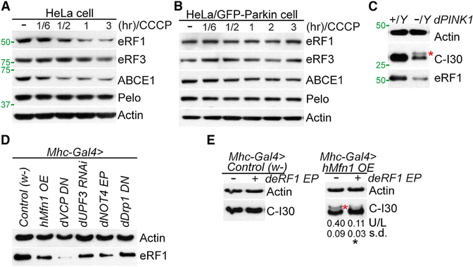
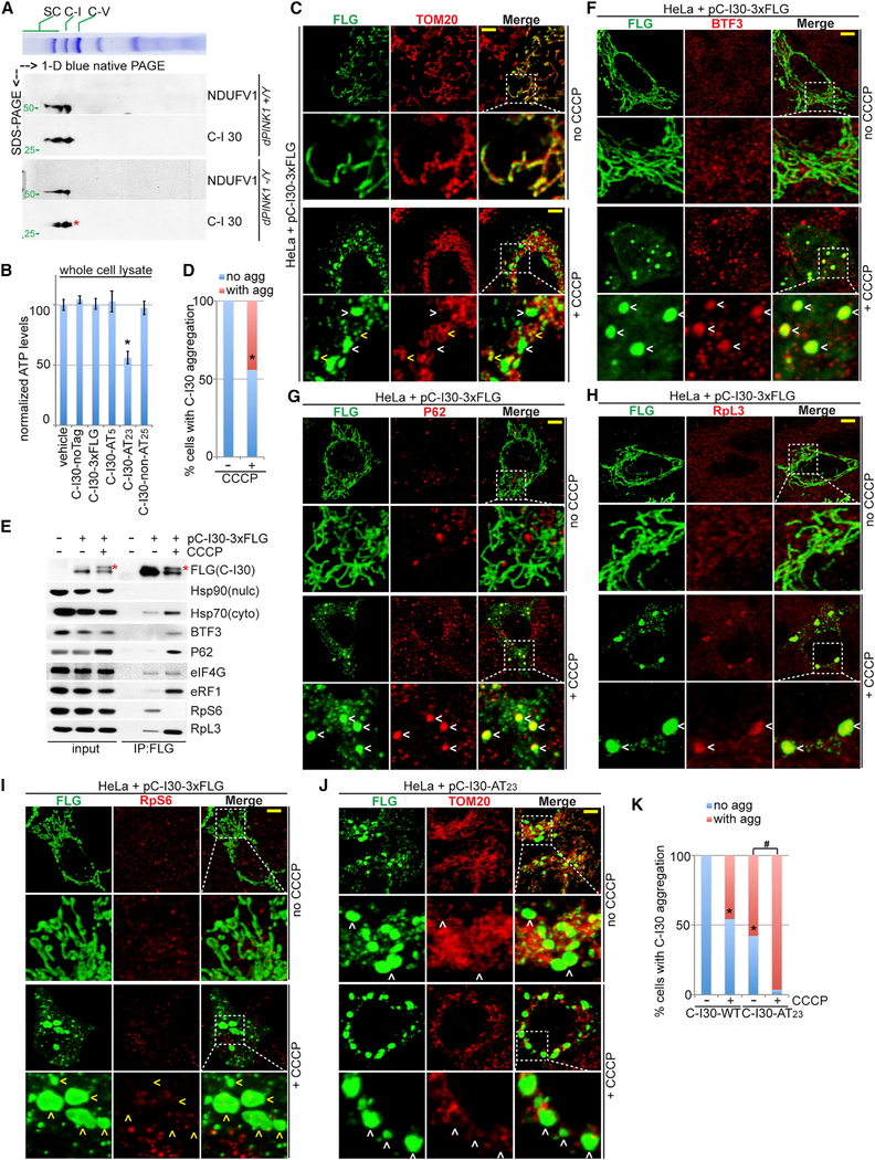
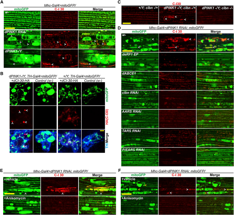
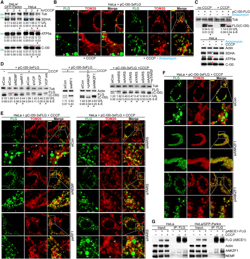
Mitochondrial dysfunction and proteostasis failure frequently coexist as hallmarks of neurodegenerative disease. How these pathologies are related is not well understood. Here, we describe a phenomenon termed MISTERMINATE (mitochondrial-stress-induced translational termination impairment and protein carboxyl terminal extension), which mechanistically links mitochondrial dysfunction with proteostasis failure. We show that mitochondrial dysfunction impairs translational termination of nuclear-encoded mitochondrial mRNAs, including complex-I 30kD subunit (C-I30) mRNA, occurring on the mitochondrial surface in Drosophila and mammalian cells. Ribosomes stalled at the normal stop codon continue to add to the C terminus of C-I30 certain amino acids non-coded by mRNA template. C-terminally extended C-I30 is toxic when assembled into C-I and forms aggregates in the cytosol. Enhancing co-translational quality control prevents C-I30 C-terminal extension and rescues mitochondrial and neuromuscular degeneration in a Parkinson's disease model. These findings emphasize the importance of efficient translation termination and reveal unexpected link between mitochondrial health and proteome homeostasis mediated by MISTERMINATE.

Keywords: CAT-tailing; MISTERMINATE; PINK1/Parkin; Parkinson’s disease; RQC; mitochondrial stress; neurodegeneration; proteostasis; ribosome stalling; translation termination.


Conflict of interest statement

DECLARATION OF INTERESTS

The authors declare no competing interests.

Figures

Figure 1.
Figure 1.. C-I30-u Formation in Mutant Flies Suffering Mitochondrial Insults
(A) Immunoblots of thoracic muscle extracts from WT (dPINK1+/Y) and PINK1 (dPINK1-/Y) flies. Asterisk indicates C-I30-u. Values under the blots represent mean and SD of the ratio of C-I30 upper band/lower band (U/L), accompanied by statistical tests, in this and all subsequent figures. Actin serves as a loading control. (B) Immunoblots of muscle samples from WT and PINK1 flies with C-I30 knocked down by two different transgenes. (C) Immunoblots of muscle samples from dParkin1/+ and dParkin1/Δ21 flies. (D) Immunoblots of muscle samples from WT and PINK1 flies with or without CCCP or rotenone (Rtn) treatment. (E) Immunoblots of muscle samples from PINK1 flies with or without H2O2 treatment. (F) Immunoblots of muscle samples from various transgenic flies. w-, WT control. See also Figure S1.
Figure 2.
Figure 2.. Co-translational QC Genes Regúlate C-I30-u Formation
(A) Immunoblots of HeLa cells transfected with C-I30-FLAG and treated with 20 μM CCCP. (B and C) Immunoblots of C-I30-FLAG and C-I30-FLAG-u after phosphatase (PPTase; B) or ubiquitin specific protease 2 (USP2; C) treatment. (D) Immunoblots of lysates from HeLa or Hela (GFP-Parkin) cells transfected with C-I30-FLAG and with or without CCCP treatment. (E) Rescue of the PINK1 wing phenotype by QC factors. *p<0.05 and **p < 0.01, Student-Newman-Keuls test (SNK-test) plus Bonferroni correction versus PINK1 group at day 7 or 15, respectively. (F) Rescue of mitochondrial morphology by QC factors in PINK1 RNAi fly muscle. (G and H) Rescue of PINK1 DA neuron mitochondrial morphology (G) and number (H) by QC factors. *p < 0.05 and **p < 0.01, SNK-test plus Bonferroni correction versus control (w-). (I) Immunoblots of muscle samples from PINK1 flies expressing UAS transgenes or enhancer P-element (EP) lines driven by Mhc-Gal4. w-, WT control. (J) Data quantification of (I). **p < 0.01 in SNK-test plus Bonferroni correction versus control (w-). Error bars represent SD from three independent assays and normalized with control. Scale bars, 5 μm (F-H). Mitochondrial morphology is monitored with mito-GFP in this and all subsequent figures. See also Figure S2.
Figure 3.
Figure 3.. C-I30-u Behaves as a CTE Form of C-I30
(A) CLIP assay showing binding of specific tRNAs to Clbn in PINK1 mutant. (B) Immunoblots of muscle samples from WT and PINK1 flies with loss of one or two copies of clbn. (C) Effect of clbn on wing posture in the WT and PINK1 conditions. *p < 0.05 or #p < 0.05 in SNK-test plus Bonferroni correction versus PINK1 at day 7 or 15, respectively. (D) Effect of clbn RNAi on mitochondrial morphology in PINK1 RNAi fly muscle. (E) Rescue of DA neuron number by clbn in PINK1 flies. **p < 0.01, two-tailed Student’s t test. (F and G) Immunoblots of muscle samples from PINK1 flies expressing AARS RNAi transgenes. (G) shows a data quantification of (F). *p < 0.05 and **p < 0.01 in SNK-test plus Bonferroni correction versus control (w-). (H) Immunoblots of muscle samples from WT and PINK1 flies treated with anisomycin. (I) Immunoblots of enterokinase (EK)-treated C-I30-FLAG and C-I30-FLAG-u. The post-digestion product showed reduced FLAG reactivity due to removal of one residue from the epitope. 1× and 4× products were loaded to match the uncut sample. (J) C-terminal peptides identified from generic database searches of fly C-I30-u peptides (see Table S1). SEVGLLNGNA, read-through sequence. (K) Longer CTE peptides identified from customized pool-based searches. Amino acids (aa) in black, C-I30 CDS; aa in blue, CTE (see Table S2). Scale bars, 5 mm (D and E). See also Figure S3.
Figure 4.
Figure 4.. eRF1 Regulates C-I30-u Formation
(A and B) Immunoblots of indicated proteins in CCCP-treated HeLa cells (A) or HeLa (GFP-Parkin) cells (B). (C) Immunoblots of eRF1 in muscle sample of WT and PINK1 flies. The same blot as in Figure 1A is used. (D) Immunoblots of eRF1 in muscle samples from WT flies expressing various transgenes. (E) Immunoblots showing removal of C-I30-u by eRF1-OE. See also Figure S4.
Figure 5.
Figure 5.. C-I30-u Is Assembled into C-I and Can Form Cytosolic Aggregates
(A) Immunoblots of 2D gel showing assembly of C-I30-u into the RCC of mitochondria purified from PINK1 flies. Another C-I marker, NDUFV1, serves as a positive control. (B) ATP measurement in HeLa cells expressing non-tagged C-I30 and C-I30-CAT-Tail. *p < 0.05, SNK-test plus Bonferroni correction versus control (w-). (C) Immunostaining showing C-I30 aggregation in CCCP-treated HeLa cells. Scale bar, 3 μm. Arrowheads indicate aggregates outside (white) or inside (yellow) mitochondria. (D) Qualification of data shown in (C). *p < 0.05, chi-squared test. (E) FLAG IP from HeLa cells transfected with C-I30-FLAG, with or without CCCP treatment. (F-I) Immunostaining of HeLa cells transfected with C-I30-FLAG and treated with or without CCCP. Scale bar, 3 μm. White arrowheads indicate aggregates colocalizing with BTF3 (F), P62 (G), or RpL3 (H), and yellow arrowheads indicate no RpS6 colocalization (I). (J) Immunostaining of HeLa cells transfected with C-I30-FLAG-AT23, with or without CCCP. Arrowheads indicate extra-mitochondrial aggregates. (K) Qualification of data shown in (J). *p < 0.05 and #p < 0.05, chi-squared test. More than 100 transfected cells in each group were quantified in (D) and (K). Scale bars, 3 μm (C, F-I, and J). See also Figure S5.
Figure 6.
Figure 6.. C-I30-u Forms Aggregates in Fly Neuromuscular Tissues
(A) Immunostaining showing C-I30 aggregates in PINK1 muscle. (B) Immunostaining showing HA+ aggregates in PINK1 DA neurons expressing C-I30-HA. (C) Immunostaining showing effect of clbn on C-I30 aggregation in PINK1 muscle. (D) Immunostaining showing effect of genetic manipulations on mitochondrial morphology and C-I30 aggregation in PINK1 RNAi fly muscle. (E and F) Short-term (1 day; E) or long-term (7 days; F) anisomycin treatment on C-I30 aggregation and mitochondrial morphology in PINK1 RNAi fly muscle. Scale bars, 5 μm. Arrowheads indicate CI-30 aggregates. See also Figure S6.
Figure 7.
Figure 7.. Mechanism of C-I30-u Formation and Aggregation in Human Cells
(A) Immunoblots showing induction of C-I30-U and ATP5a-u (red asterisk) in HeLa, but not HeLa (GFP-Parkin), cells. Blue asterisk indicates a succinate dehydrogenase subunit A (SDHA) form not responding to CCCP or Parkin and is likely SDHA preprotein. (B) Immunostaining showing effect of anisomycin on CCCP-induced C-I30-FLAG aggregation. Arrowheads indicate aggregates outside (white) or inside (yellow) mitochondria. (C) Immunoblots showing the effect of anisomycin on the CTE (red asterisk) of C-I30-FLAG (upper panel) or endogenous ATP5a and C-I30 (lower panel). Blue asterisk indicates SDHA preprotein not responding to anisomycin. (D) Immunoblots showing effects of various modifiers or VCP inhibitor on C-I30-FLAG-u levels. (E) Immunostaining showing effects of various modifiers on CCCP-induced FLAG+ aggregates. Arrowheads indicate aggregates. (F) Immunostaining showing effects of ANKZF1 RNAi or OE on C-I30-FLAG aggregation. (G) CoIP assay showing effect of Parkin on the interaction of ANKZF1 or NEMF with ABCE1. Scale bars, 3 mm (B, E, and F). See also Figure S7.

Similar articles

Cited by

References

    1. Alhebshi A, Sideri TC, Holland SL, and Avery SV (2012). The essential iron-sulfur protein Rli1 is an important target accounting for inhibition of cell growth by reactive oxygen species. Mol. Biol. Cell 23, 3582–3590. - PMC - PubMed
    1. Bi X, Jones T, Abbasi F, Lee H, Stultz B, Hursh DA, and Mortin MA (2005). Drosophila caliban, a nuclear export mediator, can function as a tumor suppressor in human lung cancer cells. Oncogene 24, 8229–8239. - PubMed
    1. Bossy-Wetzel E, Schwarzenbacher R, and Lipton SA (2004). Molecular pathways to neurodegeneration. Nat. Med 10 (Suppl), S2–S9. - PubMed
    1. Brandman O, and Hegde RS (2016). Ribosome-associated protein quality control. Nat. Struct. Mol. Biol 23, 7–15. - PMC - PubMed
    1. Brandman O, Stewart-Ornstein J, Wong D, Larson A, Williams CC, Li GW, Zhou S, King D, Shen PS, Weibezahn J, et al. (2012). A ribosome-bound quality control complex triggers degradation of nascent peptides and signals translation stress. Cell 151, 1042–1054. - PMC - PubMed

Publication types

MeSH terms