. 2019 Sep 26;47(17):8927-8940.
doi: 10.1093/nar/gkz652.

CtIP is essential for telomere replication

Affiliations

CtIP is essential for telomere replication

Susanna Stroik et al. Nucleic Acids Res. .

Abstract

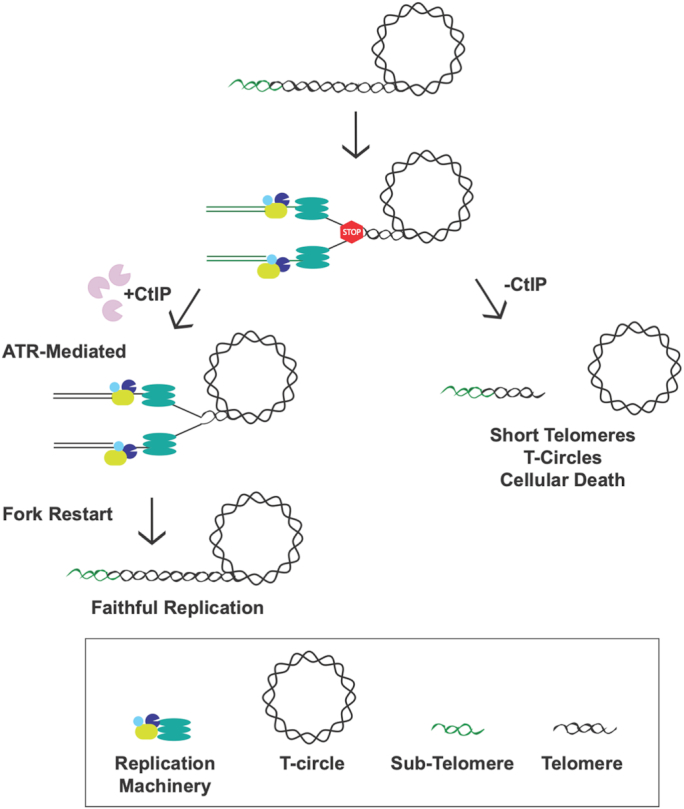
The maintenance of telomere length is critical to longevity and survival. Specifically, the failure to properly replicate, resect, and/or form appropriate telomeric structures drives telomere shortening and, in turn, genomic instability. The endonuclease CtIP is a DNA repair protein that is well-known to promote genome stability through the resection of endogenous DNA double-stranded breaks. Here, we describe a novel role for CtIP. We show that in the absence of CtIP, human telomeres shorten rapidly to non-viable lengths. This telomere dysfunction results in an accumulation of fusions, breaks, and frank telomere loss. Additionally, CtIP suppresses the generation of circular, extrachromosomal telomeric DNA. These latter structures appear to arise from arrested DNA replication forks that accumulate in the absence of CtIP. Hence, CtIP is required for faithful replication through telomeres via its roles at stalled replication tracts. Our findings demonstrate a new role for CtIP as a protector of human telomere integrity.


Figures

Figure 1.
Figure 1.
(A) A CtIP conditionally-null cell line was generated in an RPE1 background using CRISPR/Cas9 technology (scissors). LoxP sites, represented as purple triangles, were knocked-in to flank exon 2 of CtIP. The second allele was knocked out by creating a frameshift mutation (lightning bolt). CreER was stably integrated and then used to conditionally recombine out the remaining CtIP allele creating a CtIP-null cell line. (B) Confirmation of a CtIP-null cell line and a complemented derivative by western analysis. The CtIP expression levels in the indicated experimental cell lines was assessed by western blot analysis. This analysis also validated the complementation of the CtIPf/- cell line with a CtIP cDNA. GAPDH was used as a loading control. (C) CtIP-deficient cells are proliferation defective. Growth analyses of RPE1 WT, CtIPf/−, CtIPf/- cells treated with 4-OHT for 2 days and CtIPf/-:CtIP over a period of 6 days. The data shown represent the average of three biological replicates. (D) CtIP-null cells show high levels of genomic instability. Representative images of intact nuclei, micronuclei, and fragmented nuclei from CtIPf/- cells treated with 4-OHT for 3 days. Nuclei were DAPI stained; scalebar = 30 μm. (E) Quantification of the number of micronuclei observed in CtIP-null cells. 150 cells similar to the ones shown in panel (D) were scored for the presence of micronuclei in each background.
Figure 2.
Figure 2.
(A) CtIP-null cells accumulate markers of DNA damage. Representative images of RPE1 WT, CtIPf/−, or CtIPf/− cells treated with 4-OHT for 2 days, and then stained for γ-H2AX. Scale bars, 30 μm. (B) Quantification of γ-H2AX foci per cell. Data from three independent experiments are shown, n = 150, paired t-test, P < .0001 = ****. (C) Gross chromosomal abnormalities are frequently observed in metaphase-arrested CtIP-null cells. Representative images of metaphase spreads from RPE1 WT, CtIPf/−, CtIPf/− treated with 4-OHT for 2 days, or CtIPf/-:CtIP. Scale bars, 45 μm. (D) Presence of radial chromosomes, fusions and chromosomal fragments in CtIP-null cells. The boxed representative abnormalities in CtIPf/− cells treated with 4-OHT from (C) are shown in expanded form. (E) Quantification of chromosomal breaks, fusions, and radials. Data from 3 independent experiments, n = 2200, one-way ANOVA statistical analysis and Tukey's test, P < .01 = **, P < .001 = ***.
Figure 3.
Figure 3.
(A) IF analysis of telomere-associated damage in CtIP-null cells. TIFs in RPE1 WT, CtIPf/−, CtIPf/− treated with 4-OHT for 2 days, and CtIPf/-:CtIP cells were determined. Telomeres were labeled with a PNA probe and formula image-H2AX foci were visualized by IF staining. Scale bar in the bottom right panel is 10 μm, n = 300. (B) TIFs are highly elevated in CtIP-null cells. Quantification of TIFs in RPE1 WT, CtIPf/− untreated, CtIPf/- cells treated for 3 days with 4-OHT, and CtIPf/-:CtIP cells as shown in panel (A). The data are derived from three biological replicates; one-way ANOVA and Tukey's test, P < .001 = ***. (C) Telomere restriction fragment (TRF) analysis of RPE1 WT, CtIPf/−, and CtIPf/− cells treated with 4-OHT for 2 days. (D) Quantification of telomere length measurements from 3 independent TRF experiments such as shown in panel (C). Paired t-test, P < .05 = *, P < .0001 = ****. (E) Native G-overhang analysis using a 32P-radiolabeled (A2TC3)3 probe of DNA extracted from RPE1 WT, CtIPf/− untreated, and CtIPf/- cells 2 days after 4-OHT treatment. –Exo1 and +Exo1 refer to pre-treatment of the samples without or with, respectively, bacterial Exo1. (F) Quantification of G-overhang length measurements such as shown in panel (E) from 3 independent experiments. Paired t-test, P < .05 = *, P < .01 = **.
Figure 4.
Figure 4.
(A) Representative images of Telomere-FISH (T-FISH) from RPE1 WT, CtIPf/−, CtIPf/− + 4-OHT 2 days and CtIPf/-:CtIP using a (T2AG3)3 PNA telomere probe (red), as well as a AT2CGT2G2A3CG3A PNA centromere probe (green). Scale bars are 20 μm. (B) Representative images of sister chromatid fusions, chromosome:chromosome fusions, and internalized telomere signals. (C) Quantification of the telomere abnormalities such as those shown in panel (B). n = 2000, one-way ANOVA statistical test and Tukey’s test, P < .01 = **. (D) Representative images of signal free ends, centromere breaks, and breaks containing a telomere signal. (E) Quantification of the telomere abnormalities such as those shown in panel (D). The data from three independent experiments is shown. n = 2,000, one-way ANOVA and Tukey’s test, P < .01 = **, P < .001 = ***. (F) Quantification of total telomere abnormalities in CtIPf/- cells after treatment with 4-OHT for 36 h and Mirin or ATRN-119 for 24 h. The telomere aberrations scored include: signal free ends, fusions, internalized telomeres, and breaks including telomere signals. The data shown is from three independent experiments, n = 1750; one-way ANOVA and Tukey's test were used with P < .05 = *. (G) Quantification of γ-H2AX foci in CtIPf/- cells after treatment with 4-OHT for 36 h and Mirin or ATRi for 24 h. n = 250; one-way ANOVA and Tukey’s test were used, P < .01 = **.
Figure 5.
Figure 5.
(A) 2D gel analysis of telomeric DNA extracted from U-2 OS (positive control), RPE1 WT, CtIPf/-, and CtIPf/- treated with 4-OHT for 2 days. (B) Western blot analysis to confirm the siCtIP efficiency in U-2 OS cells. Actin was used as a loading control. (C) Dot blot analysis of Phi29-amplified (+Phi) or non-amplified (–Phi) c-circles from U-2 OS WT, U-2 OS +scrambled siRNA, and U-2 OS +siCtIP cells. Amplification without DNA (–DNA) served as a negative control. (D) Quantification of c-circles in U-2 OS WT, U-2 OS +scrambled siRNA, and U-2 OS +siCtIP such as those shown in panel (C) from three independent experiments. Paired t-test, P < .001 = ***.
Figure 6.
Figure 6.
(A) Representative images of arrested, partially replicated, and completely replicated telomeres in HeLa LT cells depleted of CtIP for 2 days. Replication tracts (IdU) are displayed in blue and the telomere (Telo) tracts are displayed in purple. The scale bar is 20 kb. (B) Western blot analysis confirms the siCtIP knockdown efficacy in HeLa LT cells. Cells were left untreated or treated with siRNAs (scrambled or siCtIP) and then harvested 2 days post-transfection for immunoblot analysis. (C) Quantification of telomere replication forks that were arrested, partially replicated, or completely replicated as shown in panel (A). n = 225 telomere fibers from 3 biological replicates; one-way ANOVA and Tukey's test, P < .05 = *, P < .01 = **. (D) Quantification of telomeres that were not replicated during the interval of the analysis for panel (A) in HeLa LT, HeLa LT +scrambled siRNA, and HeLa LT +siCtIP siRNA treated cells.
Figure 7.
Figure 7.
The replication fork machinery frequently stalls (STOP sign) when it encounters the telomeric sequence due to the heterochromatic nature, secondary structures, and telomere-bound proteins present. CtIP safeguards replication by facilitating either repair, restart, and/or rescue of these stalled forks leading to faithful, complete replication through the telomere. In the absence of CtIP, stalled forks persist in the telomere region eventually leading to fork collapse. Collapsed forks commonly lead to DSBs that may result in the loss of the t-loop, which, in turn, generates radically shortened telomeres and t-circles in just a few cell cycles.

Similar articles

Cited by

References

    1. de Lange T. Shelterin-mediated telomere protection. Annu. Rev. Genet. 2018; 52:223–247. - PubMed
    1. Griffith J.D., Comeau L., Rosenfield S., Stansel R.M., Bianchi A., Moss H., de Lange T.. Mammalian telomeres end in a large duplex loop. Cell. 1999; 97:503–514. - PubMed
    1. Harley C.B., Futcher A.B., Greider C.W.. Telomeres shorten during ageing of human fibroblasts. Nature. 1990; 345:458–460. - PubMed
    1. Bertuch A.A. The molecular genetics of the telomere biology disorders. RNA Biol. 2016; 13:696–706. - PMC - PubMed
    1. Baird D.M., Hendrickson E.A.. Telomeres and chromosomal translocations: there's a ligase at the end of the translocation. Adv. Exp. Med. Biol. 2018; 1044:89–112. - PubMed

Publication types