. 2019 Nov;593(21):3015-3028.
doi: 10.1002/1873-3468.13561. Epub 2019 Aug 16.

PKC induces release of a functional ectodomain of the guidance cue semaphorin6A

Affiliations

PKC induces release of a functional ectodomain of the guidance cue semaphorin6A

Riley M St Clair et al. FEBS Lett. 2019 Nov.

Abstract

Semaphorins (Semas) are a family of secreted and transmembrane proteins that play critical roles in development. Interestingly, several vertebrate transmembrane Sema classes are capable of producing functional soluble ectodomains. However, little is known of soluble Sema6 ectodomains in the nervous system. Herein, we show that the soluble Sema6A ectodomain, sSema6A, exhibits natural and protein kinase C (PKC)-induced release. We show that PKC mediates Sema6A phosphorylation at specific sites and while this phosphorylation is not the primary mechanism regulating sSema6A production, we found that the intracellular domain confers resistance to ectodomain release. Finally, sSema6A is functional as it promotes the cohesion of zebrafish early eye field explants. This suggests that in addition to its canonical contact-mediated functions, Sema6A may have regulated, long-range, forward-signaling capacity.

Keywords: PKC; eye development; guidance cue; semaphorin; semaphorin6A; zebrafish.


Figures

Figure 1.
Figure 1.. Semaphorin ectodomain peptides identified in Zappaterra et al. in human and rat embryonic cerebrospinal fluid.
A. Schematic illustrating the semaphorin (Sema) proteins that we previously identified in human and rat embryonic cerebrospinal fluid (eCSF). Asterisks indicate positions of peptides discovered in rat CSF while triangles indicate positions of peptides discovered in human CSF. Of note, all peptides identified were in the ectodomains. B. Table summarizing the number of peptides identified in each sample. If a semaphorin protein is not listed, it was not identified. C-D. HEK293 cells (C) and N2A cells (D) cells overexpressing an expression construct encoding human Sema6A were incubated in OPTI-MEM reduced serum media overnight. The conditioned media was collected and cells were lysed. Samples were run on SDS-PAGE and immunoblotted with ⍺-Sema6A. A smaller protein of approximately 95 kDa was detected in the conditioned media (arrowhead) of cells expressing full-length (FL) Sema6A (arrow).
Figure 2.
Figure 2.. Mass spectrometry analysis reveals the Sema6A ectodomain as the soluble Sema6A product.
HEK cells were transfected with an expression plasmid encoding Myc-SEMA6A. Immunoprecipitation with α-Myc was used to purify the full-length cell-bound Sema6A, and the soluble Sema6A contained in the media. A. Whole cell extracts and a small portion of the immune complexes were subjected to SDS-PAGE and western blotting to verify SEMA6A expression and a successful immunoprecipitation. Arrows indicate the full length protein and arrowheads indicate the smaller soluble Sema6A form. The multiple bands of Sema6A may indicate post-translational modifications such as glycosylation. B. The major portion of the immune complexes was subjected to coomassie staining. Each sample lane was excised according to molecular weight, digested with trypsin, and prepared for tandem mass spectrometry. C. The tryptic peptides from bands corresponding to a molecular weight of the soluble Sema6A variant, approximately 85–110 kDa, were analyzed and mapped onto human Sema6A. Gray bars indicate the location and spectral count of the tryptic peptides identified. The starred bar indicates the non-tryptic peptide detected which is predicted to be the cleavage site that produces sSema6A. As peptide coverage is negatively affected by glycosylation, we also mapped potential glycosylation sites. The letters “O” and “N” indicate predicted O- or N-linked glycosylation sites, respectively, that were identified from at least two independent glycosylation prediction programs.
Figure 3.
Figure 3.. Naturally released sSema6A can be regulated by PKC.
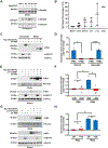
HEK293 cells were transfected with an expression construct encoding mouse Myc-Sema6A. Following overnight growth, HEK293 cells were incubated in OPTI-MEM reduced-serum media for the indicated amount of time or 6 hours if not indicated. The conditioned media was then collected and the cells were lysed. Proteins were concentrated from the conditioned media using TCA precipitation. Protein samples were subjected to SDS-PAGE and immunoblotting with the indicated antibodies. A. The time course of detectable sSema6A production, which was normalized to 18 hours, quantified in B. As sSema6A was consistently detected at 6 hours, this time point was selected for subsequent pharmacological experiments. C. Sema6A-expressing cells were treated with the PKC activator PMA (100 nM, 30 minutes) or DMSO vehicle alone during the last 30 minutes of the 6-hour conditioned media incubation. Activated Erk levels, as indicated by α-pErk, were used as a readout for PKC activity. D. Full-length (FL) Sema6A observed in the cell extract was normalized to the Sema6A PMA treated group and the Log2 fold change was quantified (left, p < 0.012). The Log2 fold change in sSema6A observed in the conditioned media was normalized to the Sema6A untreated group (right, p < 0.0001). E. PMA-induced sSema6A production is due to PKC activation. Sema6A-expressing cells were pre-treated with the selective PKC inhibitor BIM1 (5 μM) for 30 minutes prior to PMA stimulation. Log2 fold change sSema6A levels were normalized and quantified in F (+PMA/-BIM1 compared to +PMA/+BIM1 p < 0.018; -PMA/+BIM1 compared to +PMA/-BIM1 p < 0.039). G. PMA/PKC-induced sSema6A production is independent of the Mek/Erk pathway. Sema6A-expressing cells were pre-treated with the Mek inhibitor U0126 (10 μM) for 30 minutes prior to PMA stimulation. Log2 fold change sSema6A levels were normalized and quantified in H (p < 0.012). Histograms show the average of three independent experiments.
Figure 4.
Figure 4.. PKC-induced phosphorylation of Sema6A.
A-B. HEK293 cells expressing Myc-Sema6A were stimulated with PMA or DMSO vehicle control, with or without pre-treatment of BIM1. Sema6A was purified via immunoprecipitation with α-Myc and immune complexes were subjected to SDS-PAGE and immunoblotting with a PKC/AGC kinase phospho-motif specific antibody, α-RXXpS/T. A. Untreated cells show baseline RXXpS/T Sema6A phosphorylation while PMA stimulation increases this phosphorylation. BIM1 abolishes the baseline and PMA-induced phosphorylation, as quantified in B. Phosphorylation levels and total protein levels were normalized to Sema6A -PMA/-BIM1 and the Log2 fold change was calculated using the fraction phosphorylated/total Sema6A (-PMA/-BIM1 compared to –PMA/+BIM1 p < 0.0087; +PMA/-BIM1 compared to +PMA/+BIM1 p < 0.0042). Dashed line indicates cropping together due to a tear in the gel. See Figure 3E for whole cell extract Myc-Sema6A expression levels and RXXpS/T protein levels. C-E. HEK293 cells expressing Myc-Sema6A-FL or a non-phosphorylatable mutant S/T9A were stimulated with PMA or DMSO vehicle control. Sema6A was purified from the cell extract using α-Myc and RXXpS/T phosphorylation levels were measured and quantified in E (p < 0.021). The Log2 fold change in phosphorylation levels were normalized and calculated as in B. The conditioned media of Myc-Sema6A-FL- or S/T9A- expressing cells was collected and proteins were concentrated using TCA precipitation. The asterisk represents a background band in the conditioned media. The Log2 fold change in sSema6A observed in the conditioned media was normalized to the Sema6A untreated group and quantified in D. (sSema6A-FL -PMA compared to sSema6A-FL +PMA p < 0.039; sSema6A-S/T9A -PMA compared to sSema6A-S/T9A +PMA p < 0.0096). F-G. HEK293 cells expressing either Sema6A-FL or a truncated Sema6A mutant lacking the intracellular domain (Sema6A-Δcyt) were lysed and the conditioned media was collected and subjected to TCA precipitation. The Sema6A levels in the cell extract and sSema6A levels in the conditioned media were detected by immunoblotting using α-Myc. Levels were normalized to the Sema6A-FL control and the Log2 fold change was quantified in G. While there was a significant decrease in cell extract level of Sema6A-Δcyt, the truncated mutant still produced comparable levels of sSema6A (p < 0.001). Histograms show the average of two-four independent experiments.
Figure 5.
Figure 5.. Naturally released sSema6A promotes cellular cohesion of early eye fields.
A. Eyes were dissected from 18 hpf embryos of rx3:GFP transgenic zebrafish, which have GFP-positive retinal precursor cells. Individual eye explants were cultured in individual wells of a 96-well plate containing conditioned media from either untransfected or Sema6A-expressing cells for 6 hours. B-E. After incubations in conditioned media, brightfield (B, C) and fluorescent (D, E) images of the eye explants were taken and eye field cohesion was assessed by counting the number of GFP-positive ectopic cells, quantified in F, n=26 embryos incubated in control media and n=23 embryos incubated in sSema6A media, p = 0.009. Scale bar represents 75 μm. G. Cell extracts and conditioned media of untransfected or Sema6A-expressing cells were subjected to SDS-PAGE and immunoblotted with α-Sema6A to verify protein expression and the presence of sSema6A in the conditioned media.
Figure 6.
Figure 6.. Working model of Sema6A-PlxnA bidirectional signaling.
This schematic illustrates various signaling paradigms resulting from the Sema6A-PlxnA interaction. When Sema6A does not bind to PlxnA receptors, PlxnA is in an autoinhibitory conformation and forward signaling does not occur. When transmembrane Sema6A binds to PlxnA, bidirectional signal can occur, with forward signaling resulting in cellular processes such as integrin and cytoskeletal regulation, transcriptional regulation, as well as proliferation, differentiation and apoptosis. Less is known about the mechanisms governing reverse signaling, however, it is hypothesized that integrins and cytoskeletal dynamics are regulated. The ectodomain of Sema6A can be naturally released or via PKC activation, resulting in a functional soluble product, sSema6A. This would enable forward signaling with the absence of reverse signaling and represents one mechanism to regulate the direction of signaling. Interestingly, we observed sSema6A to exist as a monomer (Supplemental Figure 8). Semaphorins are classically thought to require dimerization to function, however, recombinant Sema6A ectodomains have been shown to be monomers and capable of signaling. Thus future work will investigate the binding interaction of sSema6A to PlxnA receptors.

References

    1. Luo Y, Raible D & Raper JA (1993) Collapsin: a protein in brain that induces the collapse and paralysis of neuronal growth cones, Cell. 75, 217–27. - PubMed
    1. Kolodkin AL (1996) Semaphorins: mediators of repulsive growth cone guidance, Trends Cell Biol. 6, 15–22. - PubMed
    1. Jongbloets BC & Pasterkamp RJ (2014) Semaphorin signalling during development, Development. 141, 3292–7. - PubMed
    1. Kruger RP, Aurandt J & Guan KL (2005) Semaphorins command cells to move, Nat Rev Mol Cell Biol. 6, 789–800. - PubMed
    1. Treps L, Le Guelte A & Gavard J (2013) Emerging roles of Semaphorins in the regulation of epithelial and endothelial junctions, Tissue Barriers. 1, e23272. - PMC - PubMed

Publication types

MeSH terms

LinkOut - more resources