. 2019 Oct;176(20):4065-4078.
doi: 10.1111/bph.14821. Epub 2019 Oct 10.

The ellagitannin metabolite urolithin C is a glucose-dependent regulator of insulin secretion through activation of L-type calcium channels

Affiliations

The ellagitannin metabolite urolithin C is a glucose-dependent regulator of insulin secretion through activation of L-type calcium channels

Morgane Bayle et al. Br J Pharmacol. 2019 Oct.

Abstract

Background and purpose: The pharmacology of polyphenol metabolites on beta-cell function is largely undetermined. We sought to identify polyphenol metabolites that enhance the insulin-secreting function of beta-cells and to explore the underlying mechanisms.

Experimental approach: INS-1 beta-cells and rat isolated islets of Langerhans or perfused pancreas preparations were used for insulin secretion experiments. Molecular modelling, intracellular Ca2+ monitoring, and whole-cell patch-clamp recordings were used for mechanistic studies.

Key results: Among a set of polyphenol metabolites, we found that exposure of INS-1 beta-cells to urolithins A and C enhanced glucose-stimulated insulin secretion. We further characterized the activity of urolithin C and its pharmacological mechanism. Urolithin C glucose-dependently enhanced insulin secretion in isolated islets of Langerhans and perfused pancreas preparations. In the latter, enhancement was reversible when glucose was lowered from a stimulating to a non-stimulating concentration. Molecular modelling suggested that urolithin C could dock into the Cav 1.2 L-type Ca2+ channel. Calcium monitoring indicated that urolithin C had no effect on basal intracellular Ca2+ but enhanced depolarization-induced increase in intracellular Ca2+ in INS-1 cells and dispersed cells isolated from islets. Electrophysiology studies indicated that urolithin C dose-dependently enhanced the L-type Ca2+ current for levels of depolarization above threshold and shifted its voltage-dependent activation towards more negative potentials in INS-1 cells.

Conclusion and implications: Urolithin C is a glucose-dependent activator of insulin secretion acting by facilitating L-type Ca2+ channel opening and Ca2+ influx into pancreatic beta-cells. Our work paves the way for the design of polyphenol metabolite-inspired compounds aimed at ameliorating beta-cell function.

Background and Purpose: The pharmacology of polyphenol metabolites on beta‐cell function is largely undetermined. We sought to identify polyphenol metabolites that enhance the insulin‐secreting function of beta‐cells and to explore the underlying mechanisms.

Experimental Approach: INS‐1 beta‐cells and rat isolated islets of Langerhans or perfused pancreas preparations were used for insulin secretion experiments. Molecular modelling, intracellular Ca2+ monitoring, and whole‐cell patch‐clamp recordings were used for mechanistic studies.

Key Results: Among a set of polyphenol metabolites, we found that exposure of INS‐1 beta‐cells to urolithins A and C enhanced glucose‐stimulated insulin secretion. We further characterized the activity of urolithin C and its pharmacological mechanism. Urolithin C glucose‐dependently enhanced insulin secretion in isolated islets of Langerhans and perfused pancreas preparations. In the latter, enhancement was reversible when glucose was lowered from a stimulating to a non‐stimulating concentration. Molecular modelling suggested that urolithin C could dock into the Cav1.2 L‐type Ca2+ channel. Calcium monitoring indicated that urolithin C had no effect on basal intracellular Ca2+ but enhanced depolarization‐induced increase in intracellular Ca2+ in INS‐1 cells and dispersed cells isolated from islets. Electrophysiology studies indicated that urolithin C dose‐dependently enhanced the L‐type Ca2+ current for levels of depolarization above threshold and shifted its voltage‐dependent activation towards more negative potentials in INS‐1 cells.

Conclusion and Implications: Urolithin C is a glucose‐dependent activator of insulin secretion acting by facilitating L‐type Ca2+ channel opening and Ca2+ influx into pancreatic beta‐cells. Our work paves the way for the design of polyphenol metabolite‐inspired compounds aimed at ameliorating beta‐cell function.


Conflict of interest statement

The authors declare no conflicts of interest.

Figures

Figure 1
Figure 1
Effects of urolithins A, B, C, and D on basal and glucose‐induced insulin secretion in INS‐1 cells. The effects of increasing concentrations (2–20 μmol·L−1) of (a) urolithin A, (b) urolithin B, (c) urolithin C, and (d) urolithin D in INS‐1 cells were determined under basal non‐stimulating (1.4 mmol·L−1 glucose) or 8.3 mmol·L−1 glucose‐stimulated conditions. Inserts depict the correlation between insulin secretion and the log of urolithin concentration. Under non‐stimulating glucose condition, there was a significant effect of the treatment for urolithin B and C. Under 8.3 mmol·L−1 glucose‐stimulated condition, there was a significant effect of the treatment for urolithins A and C. *P < .05, significantly different from 0 μmol·L−1 urolithin, one‐way ANOVA with Holm–Sidak test, n = 5
Figure 2
Figure 2
Effects of urolithins A, B, C, and D on insulin secretion induced by glibenclamide in INS‐1 cells. The effects of increasing concentrations (2–20 μmol·L−1) of urolithin A (URO‐A), urolithin B (URO‐B), urolithin C (URO‐C), and urolithin D (URO‐D) were determined in the presence of 1.4 mmol·L−1 glucose + 10 nmol·L−1 glibenclamide (+Glib). There was a significant effect of the treatment for urolithins A, C, and D. *P < .05, significantly different from 0 μmol·L−1 urolithin in the presence of glibenclamide; one‐way ANOVA with Holm–Sidak test, n = 5
Figure 3
Figure 3
Effects of urolithins A, B, C, and D on insulin secretion in dysfunctional INS‐1 cells. INS‐1 cells were cultured in a control medium (CT) or in a medium containing 25 mmol·L−1 glucose for 48 hr (glucotoxicity). Then, cells were incubated with 8.3 mmol·L−1 glucose, in the absence (Veh) or in the presence of 20 μmol·L−1 urolithin A (URO‐A), urolithin B (URO‐B), urolithin C (URO‐C), or urolithin D (URO‐D). There was a significant effect of the treatment under glucotoxicity for urolithins A and C. *P < .05, significantly different from Veh under glucotoxicity, one‐way ANOVA with Holm–Sidak test, n = 7
Figure 4
Figure 4
Effect of urolithin C on basal and glucose‐induced insulin secretion in rat pancreatic islets. Rat pancreatic islets were incubated in the absence (Vehicle) or in the presence of 20 μmol·L−1 urolithin C (URO‐C) under basal non‐stimulating (2.8 mmol·L−1 glucose) or 8.3 mmol·L−1 glucose‐stimulated conditions. There was an effect of the glucose and urolithin C as well as an interaction between both factors. *P < .05, significantly different as indicated, ns, non‐significant; two‐way ANOVA with Holm–Sidak test, n = 6
Figure 5
Figure 5
Effects of urolithin C on insulin release in rat isolated perfused pancreas. (a) A whole pancreas was isolated prior to perfusion with 8.3 mmol·L−1 glucose throughout the experiment. After the addition of urolithin C or vehicle (from 17 to 47 min) there was an effect of the treatment and an interaction between time and treatment. *P < .05 significantly different from vehicle; two‐way ANOVA with repeated measures and Holm–Sidak test. Insert depicts the mean AUC of insulin release before (0–15 min) and after (17–47 min) addition of urolithin C or vehicle. *P < .05, significantly different as indicated, ns, non‐significant; t‐test; n = 5. (b) A pancreas was perfused with 5 mmol·L−1 of glucose throughout the experiment and with urolithin C for 10 min, n = 5. (c) A pancreas was perfused with 8.3 mmol·L−1 glucose before addition of urolithin C and then glucose concentration was reduced to 5 mmol·L−1. (d) Histogram depicts the AUC of insulin release (from panel C) at different time ranges. There was a significant effect of the treatment. *P < .05, significantly different as indicated; one‐way ANOVA with Holm–Sidak test, n = 5. The arrows indicate the time point of urolithin C addition (20 μmol·L−1)
Figure 6
Figure 6
Docking of urolithin C at the Cav1.2 channel α1C subunit model. The Cav1.2 channel α1c subunit model and urolithin C are shown in green and cyan respectively. (a) Large view of urolithin C docked inside the Cav1.2 channel. (b) Zoom view of urolithin C docked inside the Cav1.2 channel showing the hydrogen bonding in black dash. (c) Schematic representation of the hydrogen bonding between urolithin C and the Cav1.2 channel
Figure 7
Figure 7
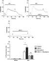
Effects of urolithin C on depolarisation‐induced rise in intracellular calcium in INS‐1 cells. [Ca2+]i was monitored using the ratiometric fluorescent Ca2+ indicator Fura‐2AM. INS‐1 cells were incubated with 20 μmol·L−1 urolithin C in the absence or presence of 20 μmol·L−1 of nimodipine. Typical recordings illustrate variations of fluorescence ratio (F340/F380) and rise in [Ca2+]i in response to 15 mmol·L−1 KCl in the absence (a) or presence of urolithin C (b) and in response to urolithin C under basal condition (c). (d) The histogram depicts the mean of the maximal variation in the fluorescence ratio. There was an effect of KCl and the treatment as well as an interaction between both factors. *P < .05, significantly different as indicated; ns, non‐significant; two‐way ANOVA with Holm–Sidak test; n = 6
Figure 8
Figure 8
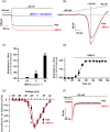
Effects of urolithin C on L‐ and T‐type Ca2+ channel currents carried by Ba2+ in INS‐1 cells. (a) Typical traces of slow inactivated L‐type Ca2+ current in the absence (Veh), presence of 20 μmol·L−1 urolithin C (URO‐C) or urolithin C + nimodipine (20 μmol·L−1). (b) Typical traces of L‐type Ca2+ current stimulated by a ramp depolarization in the absence (Veh) or presence of 20 μmol·L−1 urolithin C. (c) Concentration–response of urolithin C on the L‐type current recorded at −10 mV. The histogram depicts the mean of current expressed as the percentage of current obtained in the presence of vehicle, n = 5. (d) Time course of the effect of 20 μmol·L−1 of urolithin C on L‐type Ca2+ current (frequency of stimulation of 0.1 Hz, urolithin C was perfused at t = 20 s). The data represent the mean of current expressed as the percentage of current obtained in the presence of vehicle, n = 5. (e) Effect of 20 μmol·L−1 of urolithin C on the current‐to‐voltage (I/V) relationship of L‐type current. Both curves were normalized to 1 (maximum current; holding potential −80 mV, depolarization steps of 10‐mV increments at 0.1 Hz from −80 to 40 mV). For the negative potentials there was an effect of the treatment and an interaction between treatment and depolarization. *P < .05 significantly different from Veh at −20 mV, two‐way ANOVA with Holm–Sidak test, n = 5. (f) Effect of 20 μmol·L−1 of urolithin C on typical T‐type Ca2+ current

References

    1. Alexander, S. P. H. , Fabbro, D. , Kelly, E. , Marrion, N. V. , Peters, J. A. , Faccenda, E. , … Collaborators, C. G. T. P. (2017). The Concise Guide to PHARMACOLOGY 2017/18: Catalytic receptors. British Journal of Pharmacology, 174, S225–S271. 10.1111/bph.13876 - DOI - PMC - PubMed
    1. Alexander, S. P. H. , Striessnig, J. , Kelly, E. , Marrion, N. V. , Peters, J. A. , Faccenda, E. , … CGTP Collaborators (2017). The Concise Guide to PHARMACOLOGY 2017/18: Voltage‐gated ion channels: THE CONCISE GUIDE TO PHARMACOLOGY 2017/18: Voltage‐gated ion channels. British Journal of Pharmacology, 174, S160–S194. 10.1111/bph.13884 - DOI - PMC - PubMed
    1. Bai, X. , Yang, P. , Zhou, Q. , Cai, B. , Buist‐Homan, M. , Cheng, H. , … Shi, G. (2017). The protective effect of the natural compound hesperetin against fulminant hepatitis in vivo and in vitro. British Journal of Pharmacology, 174, 41–56. 10.1111/bph.13645 - DOI - PMC - PubMed
    1. Bardy, G. , Virsolvy, A. , Quignard, J. F. , Ravier, M. A. , Bertrand, G. , Dalle, S. , … Oiry, C. (2013). Quercetin induces insulin secretion by direct activation of L‐type calcium channels in pancreatic beta cells. British Journal of Pharmacology, 169, 1102–1113. 10.1111/bph.12194 - DOI - PMC - PubMed
    1. Bayle, M. , Roques, C. , Marion, B. , Audran, M. , Oiry, C. , Bressolle‐Gomeni, F. M. M. , & Cros, G. (2016). Development and validation of a liquid chromatography‐electrospray ionization‐tandem mass spectrometry method for the determination of urolithin C in rat plasma and its application to a pharmacokinetic study. Journal of Pharmaceutical and Biomedical Analysis, 131, 33–39. 10.1016/j.jpba.2016.07.046 - DOI - PubMed

Publication types