Review
. 2019 Sep;41(9):e1900062.
doi: 10.1002/bies.201900062. Epub 2019 Aug 5.

Half-Intercalation Stabilizes Slipped Mispairing and Explains Genome Vulnerability to Frameshift Mutagenesis by Endogenous "Molecular Bookmarks"

Affiliations
Review

Half-Intercalation Stabilizes Slipped Mispairing and Explains Genome Vulnerability to Frameshift Mutagenesis by Endogenous "Molecular Bookmarks"

Andrei Kuzminov. Bioessays. 2019 Sep.

Abstract

Some 60 years ago chemicals that intercalate between base pairs of duplex DNA were found to amplify frameshift mutagenesis. Surprisingly, the robust induction of frameshifts by intercalators still lacks a mechanistic model, leaving this classic phenomenon annoyingly intractable. A promising idea of asymmetric half-intercalation-stabilizing frameshift intermediates during DNA synthesis has never been developed into a model. Instead, researchers of frameshift mutagenesis embraced the powerful slipped-mispairing concept that unexpectedly struggled with the role of intercalators in frameshifting. It is proposed that the slipped mispairing and the half-intercalation ideas are two sides of the same coin. Further, existing findings are reviewed to test predictions of the combined "half-intercalation into the slipped-mispairing intermediate" model against accumulated knowledge. The existence of potential endogenous intercalators and the phenomenon of "DNA bookmarks" reveal ample possibilities for natural frameshift mutagenisis in the cell. From this alarming perspective, it is discussed how the cell could prevent genome deterioration from frameshift mutagenesis.

Keywords: 1 base pair (bp) indels; DNA synthesis; acridines; asymmetric intercalations; ethidium bromide; strand slippage.


Conflict of interest statement

The author declares no conflict of interest.

Figures

Fig. 1.
Fig. 1.. The early findings and models.
A. Alternative base pairing explains transition mutations. Double arrows indicate base pairing. The two natural base pairs (A:T, G:C) are shown in black. A pyrimidine base analog bromodeoxyuridine (BrdU), that can pair with either adenine or guanine, is shown in blue. A purine base analog 2-aminopurine (2AP), that can pair with either thymine or cytosine, is shown in red. B. The structure of two intercalator mutagens: 9-aminoacridine and proflavine. C. The model of Lerman of acridine interaction within duplex DNA. DNA base pairs are shown as light blue “pebbles” stacked on each other, surrounded by the two darker blue spirals of the sugar-phosphate backbone. The normal DNA duplex is shown on the left. A duplex in which four orange molecules of acridine have intercalated is shown on the right. Note that the backbone is distorted to accommodate intercalation. Also, fewer base pairs per turn means that DNA accommodating intercalators becomes positively-supercoiled. The black “X” marks the position of crossing-over between the two DNAs, according to Lerman’s explanation of how intercalation induces frameshift mutagenesis.
Fig. 2.
Fig. 2.. Brenner’s idea of half-intercalation inducing frameshift mutagenesis.
DNA structure is shown in navy, the frameshift-affected base pair is in light blue, the intercalator molecule is in red. A. Normal DNA synthesis. B. Half-intercalation in the primer strand leads to 1-nt deletion. C. Half-intercalation in the template strand leads to 1-nt insertion.
Fig. 3.
Fig. 3.. Streisinger’s model of slipped mispairing to explain frameshift mutagenesis and possible roles of intercalators in amplifying this mutagenesis (framed).
DNA structure is shown in navy, the frameshift-affected base pair (A, B and F only) is in light blue, the nucleotide that is or will be pushed extrahelical is in light purple, the intercalator molecule is in red. A. Normal DNA synthesis. B. The primer strand 3’-end slips one nucleotide backwards on a homonucleotide run, forming a compensatory bulge with the extra base bulging out. C-F: Various ideas on how intercalators could promote frameshifts. C. The slipped mispairing intermediate is stabilized by two symmetric intercalations into the DNA duplex on both sides of the bulge. D. The slipped mispairing is stabilized by stacking interactions with intercalators, sandwiching the extrahelical base. E. Slipped mispairing is caused by intercalation into the ssDNA template. F. (Not a part of the original model) Half-intercalation across the extra base absorbs the bulge into the duplex and stabilizes the slipped mispairing intermediate.
Fig. 4.
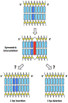
Fig. 4.. A scheme of a typical textbook “explanation” of how symmetric intercalation causes 1-bp-indels (frameshift mutations).
The two strands of the DNA duplex are shown with structural details, with bases represented by blue rectangles and deoxyriboses by yellow pentagons. The base pair affected in the frameshift mutations is shown in a darker blue, the intercalator is in red.
Fig. 5.
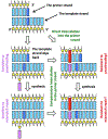
Fig. 5.. A model of 1nt-deletion caused by slipped-back template strand with subsequent compensatory half-intercalation into the primer strand.
The color designation is like in Fig. 4; in addition, the base of the extra-helical nucleotide is shown in lavender. After the template strand slips back, the generic scenario demands removal of the 3’-end of the primer if it does not match the template. The three frameshift-promoting intercalation possibilities are: 1) direct intercalation into the primer strand causing the slippage; 2) right after the slippage (still within the polymerase), potentially inhibiting proofreading; 3) after the polymerase has left, potentially inhibiting mismatch repair.
Fig. 6.
Fig. 6.. A model of 1nt-insertion caused by slipped-back primer strand with subsequent compensatory half-intercalation into the template strand.
The color designation is like in Fig. 5. In contrast to the first frameshift-promoting intercalation scenario of Fig. 5, here intercalation without prior slippage is into the single-stranded template.
Fig. 7.
Fig. 7.. Half-intercalation via wedging and bookmarks.
DNA structure is shown in navy, the intercalator is in red. A. Asymmetric intercalation should lead to “wedging” into the DNA structure. B. DNA and the bookmark tripeptide Lys-Trp-Lys. C. Due to its positive charge, the tripeptide interacts tightly with the negatively-charged DNA backbone. D. The tryptophanyl residue wedges into the DNA duplex.

Similar articles

References

    1. Avery OT, Macleod CM, McCarty M, J. Exp. Med 1944, 79, 137; A. D. Hershey, M. Chase, J. Gen. Physiol. 1952, 36, 39. - PMC - PubMed
    1. Mirsky AE, Ris H, Nature 1949, 163, 666; H. Swift, Proc. Natl. Acad. Sci. U S A 1950, 36, 643; H. H. Swift, Physiol. Zool. 1950, 23, 169. - PubMed
    1. Franklin RE, Gosling RG, Nature 1953, 171, 740; R. E. Frankling, R. G. Gosling, Nature 1953, 172, 156. - PubMed
    1. Watson JD, Crick FHC, Nature 1953, 171, 737; J. D. Watson, F. H. C. Crick, Nature 1953, 171, 964. - PubMed
    1. Hollaender A, Annu. Rev. Physiol 1946, 8, 1. - PubMed

Publication types

MeSH terms

LinkOut - more resources