. 2019 Jul 16:13:289.
doi: 10.3389/fncel.2019.00289. eCollection 2019.

Enriched Environment Shortens the Duration of Action Potentials in Cerebellar Granule Cells

Affiliations

Enriched Environment Shortens the Duration of Action Potentials in Cerebellar Granule Cells

Abdelmoneim Eshra et al. Front Cell Neurosci. .

Abstract

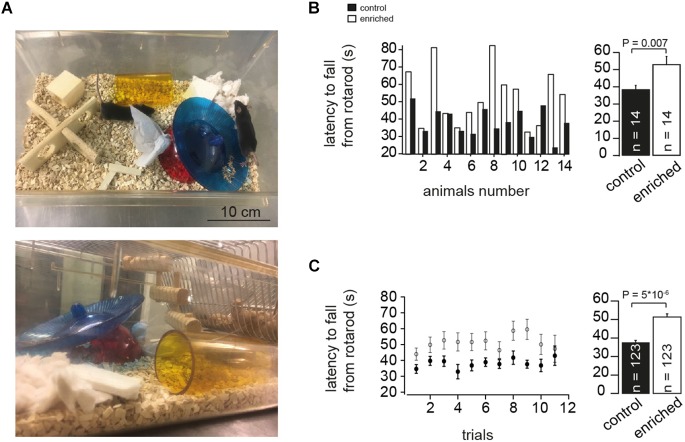
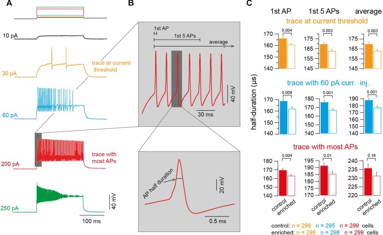
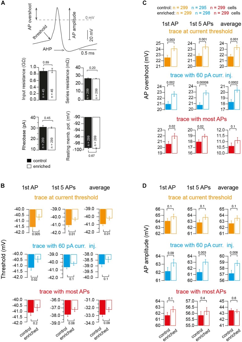
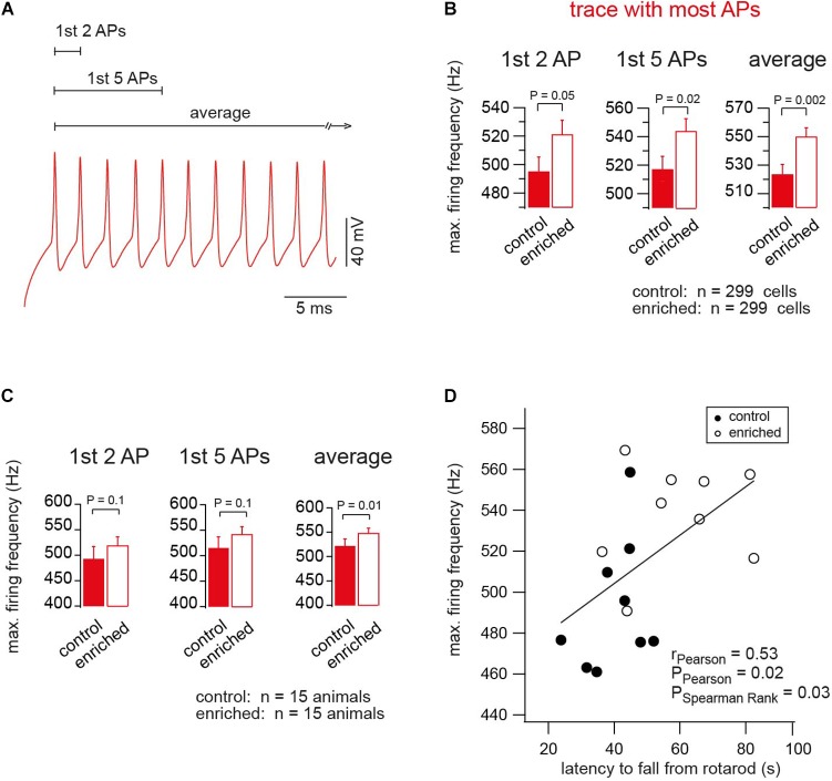
Environmental enrichment for rodents is known to enhance motor performance. Structural and molecular changes have been reported to be coupled with an enriched environment, but functional alterations of single neurons remain elusive. Here, we compared mice raised under control conditions and an enriched environment. We tested the motor performance on a rotarod and subsequently performed whole-cell patch-clamp recordings in cerebellar slices focusing on granule cells of lobule IX, which is known to receive vestibular input. Mice raised in an enriched environment were able to remain on an accelerating rotarod for a longer period of time. Electrophysiological analyses revealed normal passive properties of granule cells and a functional adaptation to the enriched environment, manifested in faster action potentials (APs) with a higher depolarized voltage threshold and larger AP overshoot. Furthermore, the maximal firing frequency of APs was higher in mice raised in an enriched environment. These data show that enriched environment causes specific alterations in the biophysical properties of neurons. Furthermore, we speculate that the ability of cerebellar granule cells to generate higher firing frequencies improves motor performance.

Keywords: action potential; cerebellum; electrophysiology; enriched environment; granule cell.


Figures

FIGURE 1
FIGURE 1
Enriched environment improves motor performance. (A) Example photographs of EE cage showing different items placed during the housing of the mice. (B) Left: Bar graphs showing comparison of the absolute latency on the rotarod per animal (animal-to-animal basis). Right: Bar graph of the average latency on the rotarod assessment test for EE and control mice, n = 14 and 14 mice, respectively; Student’s t-test was used to measure statistical significance, Pt-test = 0.007. (C) Left: Bar graphs showing comparison of the absolute latency on the rotarod per trial number (trial-to-trial basis). Right: Bar graph of the grand average of the latency on the rotarod, n = 123 and 123 trials, for control and EE mice, respectively; Pt-test = 5 × 10⁻⁶.
FIGURE 2
FIGURE 2
Action potentials of EE mice have shorter half duration. (A) Example voltage traces upon injecting current of increasing amplitudes (orange: trace at current threshold; cyan: trace with 60-pA-current injection; red: trace with maximum APs fired; green: trace with highest current injection and decrease in APs number). (B) Top: Magnification of the shaded part in panel (A) showing an example of APs. Bottom: Corresponding magnification showing a single AP. (C) Average AP half duration for control and EE mice (orange: trace at current threshold, n = 299 and 299 cells; cyan: trace with 60-pA-current injection, n = 295 and 298 cells; red: trace with maximum APs fired, n = 299 and 299 cells). From left to right, analysis of first AP, first five APs, and average of all APs. All the P-values shown are from Student’s t-test.
FIGURE 3
FIGURE 3
Alteration in threshold, overshoot and amplitude of action potentials upon enrichment. (A) Example of an AP illustrating the criteria of measuring AP threshold, AP overshoot and AP amplitude. Bar graphs showing the average of resting membrane potential, n = 299 and 299 cells; input resistance, n = 45 and 45 cells; rheobase, n = 299 and 299 cells; and series resistance, n = 299 and 299 cells, of neurons of control and EE mice, respectively. (B) Average voltage threshold of APs of neurons of control and EE mice (orange: trace at current threshold, n = 299 and 299 cells; cyan: trace with 60-pA-current injection, n = 295 and 298 cells; red: trace with maximum APs fired, n = 299 and 299 cells). From left to right, analysis of first AP, first five APs and average of all APs, respectively. (C) Corresponding average AP overshoot. (D) Corresponding average AP amplitude. All the P-values shown are from Student’s t-test.
FIGURE 4
FIGURE 4
Enriched environment tunes neurons for firing at higher frequencies. (A) An example of the APs of the trace of maximum firing frequency (color-code as in Figure 2). (B) Average maximum firing frequency of neurons of EE and control mice, n = 299, 299 cells, based on the first two, first five APs, and average of all APs in the trace, in which most APs were fired, Pt-test = 0.05, 0.02, 0.002, respectively. (C) Average maximum firing frequency of neurons of EE and the corresponding control mice, n = 15, 15 mice. For the first two, first five APs and average of all APs in the trace, in which most APs were fired, Pt-test = 0.1, 0.1 and 0.01, respectively. (D) Correlation between the maximum firing frequency and the latency of the mouse to fall from rotarod, for control and EE mice, n = 9 and 9 mice, rPearson = 0.53, PPearson = 0.02, and PSpearmanRank = 0.03.

Similar articles

Cited by

References

    1. Adrian E. D., Bronk D. W. (1929). The discharge of impulses in motor nerve fibres: part II. The frequency of discharge in reflex and voluntary contractions. J. Physiol. 67 119–151. 10.1113/jphysiol.1929.sp002557 - DOI - PMC - PubMed
    1. Arenz A., Silver R. A., Schaefer A. T., Margrie T. W. (2008). The contribution of single synapses to sensory representation in vivo. Science 321 977–980. 10.1126/science.1158391 - DOI - PMC - PubMed
    1. Barak B., Shvarts-Serebro I., Modai S., Gilam A., Okun E., Michaelson D. M., et al. (2013). Opposing actions of environmental enrichment and Alzheimer’s disease on the expression of hippocampal microRNAs in mouse models. Transl. Psychiatry 3:e304. 10.1038/tp.2013.77 - DOI - PMC - PubMed
    1. Barmack N. H. (2003). Central vestibular system: vestibular nuclei and posterior cerebellum. Brain Res. Bull. 60 511–541. 10.1016/S0361-9230(03)00055-8 - DOI - PubMed
    1. Carta M., Lanore F., Rebola N., Szabo Z., Da Silva S. V., Lourenço J., et al. (2014). Membrane lipids tune synaptic transmission by direct modulation of presynaptic potassium channels. Neuron 81 787–799. 10.1016/j.neuron.2013.12.028 - DOI - PubMed